Acute Neuromuscular Weakness

Chapter 67


Acute Neuromuscular Weakness


The development of acute neuromuscular weakness is one of the more common neurologic syndromes requiring admission to the intensive care unit (ICU). Because respiratory muscle weakness is associated with these disorders, hypercapnic respiratory failure is a frequent complication. Furthermore, bulbar weakness can lead to difficulty swallowing and weakened cough, increasing the risk of aspiration.


General Approach to Acute Neuromuscular Weakness


Monitoring for Respiratory Compromise


In the setting of neuromuscular disorders, careful respiratory monitoring should be the first priority in the care of these patients. The same general approach to respiratory failure applies to all neuromuscular disorders. Although oxygenation should be monitored closely in these patients, pulse oximetry and arterial blood gases (ABGs) cannot be relied on to gauge acute respiratory muscle weakness. This is because abnormalities in these measures frequently lag behind respiratory muscle fatigue or weakness that will imminently result in respiratory failure. Vital capacity (VC) and maximal inspiratory pressure (MIP) are more useful in these patients. The vital capacity reflects the mechanical strength of the muscles of respiration and can be easily performed at the bedside. Maximal inspiratory pressure provides similar information and, like the vital capacity, can be checked frequently at the bedside. For patients with acute neuromuscular weakness, the VC, MIP, or both should be repeated at frequent intervals. How frequently these parameters should be measured depends on the disease and the rate of progression of weakness; for example, every 2 hours may be desirable in a patient with a rapidly worsening myasthenic crisis.


In some patients it is difficult to accurately assess the VC or MIP because of facial weakness (poor seal with the mask) or delirium. Thus, these measures may be falsely low. The VC and MIP should always be interpreted in the context of the clinical signs of respiratory muscle weakness (such as increasing respiratory rate, tachycardia, use of accessory muscles of respiration, and paradoxical motion of the diaphragm), as well as the trend in these measures.


Need for Ventilatory Support


In the setting of worsening respiratory weakness or clinical concern about the patient’s ability to protect their airway, the clinician should be aggressive about elective intubation. With close monitoring, endotracheal intubation should be performed as an elective procedure before precipitous respiratory collapse (i.e., not in response to this complication). As long as the patient does not need an artificial airway to clear secretions or prevent airway occlusion, noninvasive ventilation may serve as a useful temporizing modality to provide assisted ventilation, particularly if the respiratory muscle weakness can be reversed quickly (see Chapter 3), but concern for aspiration is an important consideration in this scenario.


The following parameters warn of impending respiratory arrest and, generally, are indications for intubation (Box 67.1): vital capacity (VC) less than 20 mL/kg predicted body weight (PBW) (see footnote of Box 67.1 for formulas to calculate PBW); VC falling toward 20 mL/kg PBW plus clinical signs of respiratory muscle fatigue, such as increasing respiratory rate, tachycardia, use of accessory muscles of respiration, and paradoxical motion of the diaphragm (“respiratory paradox”); maximal inspiratory pressure (MIP) worse (i.e., less negative) than –30 cm H2O; or maximal expiratory pressure < 40 cm H2O. This general guide has been termed by some the “20-30-40 rule.” Oropharyngeal weakness with an inability to handle secretions and risk of aspiration or airway obstruction also may necessitate intubation.


Differential Diagnosis


In evaluating a patient with acutely worsening neuromuscular weakness, it is important to determine if the patient has a known history of neuromuscular disease, such as myasthenia gravis or amyotrophic lateral sclerosis (ALS). Neuromuscular respiratory failure may be part of the natural history of the disease or may be precipitated by transiently increased weakness resulting from an underlying infection, such as a urinary tract infection or pneumonia. Infectious or metabolic disorders should be thoroughly investigated and treated, if found.


Disorders that produce neuromuscular weakness severe enough to result in ICU care, such as myasthenia gravis or the Guillain-Barré syndrome, are generally separated from disorders that develop de novo in the ICU setting (the latter are discussed in Chapter 48).


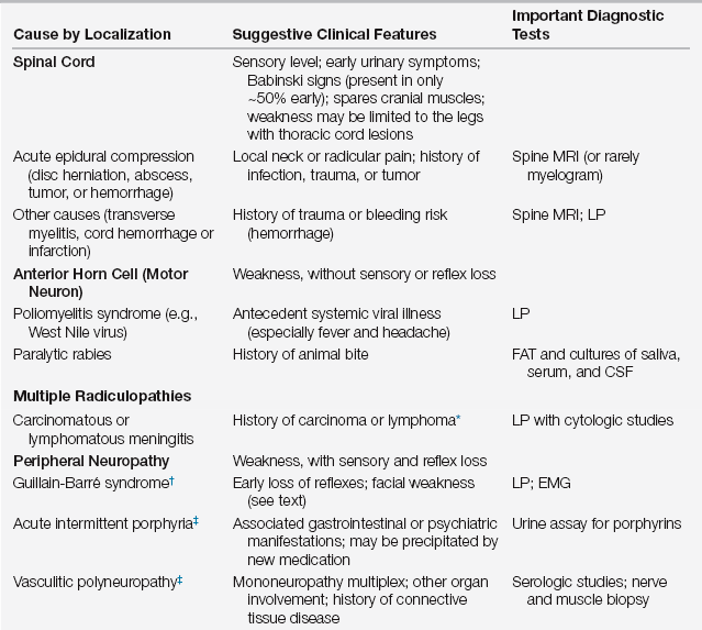
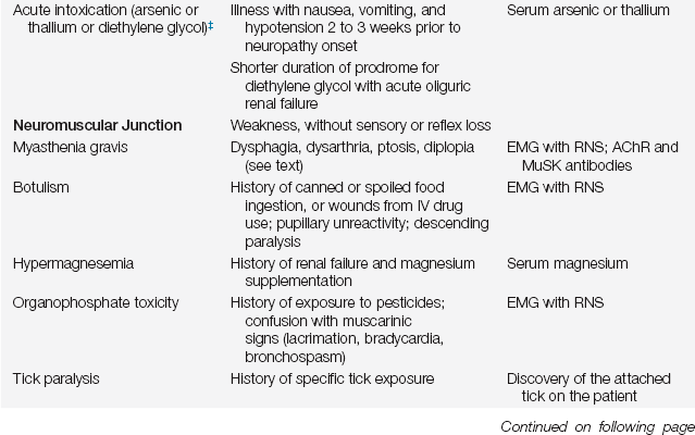
Not infrequently, the cause of the neuromuscular weakness that results in ICU care is unknown. This necessitates a diagnostic workup because pharmacologic and other interventions vary according to specific causes. In the differential diagnosis of acute neuromuscular weakness, certain clues from the history and examination can help to identify its cause (Table 67.1).


Electrophysiologic studies (nerve conduction studies and needle electromyography, which are collectively called electromyography [EMG]) are usually required to fully investigate these disorders. This is particularly true in individuals with an abnormal mental status (common in the ICU setting) who may not be able to cooperate with a motor and sensory examination. EMG allows full investigation of the presence and nature of peripheral motor and sensory involvement. It can distinguish among disorders of nerve, muscle, and neuromuscular junction. It also provides prognostic information by quantifying the extent of nerve or muscle injury.


Nerve and muscle biopsy are invasive and are not generally performed in the ICU setting, unless there is suspicion of another disorder that is only identified pathologically. Of the few indications for performing a nerve biopsy, suspected vasculitis is the most common. Muscle biopsy should likewise be reserved for patients in whom a specific cause is likely to be identified only by biopsy, such as an inflammatory myopathy (myositis).


The Guillain-Barré Syndrome


The Guillain-Barré syndrome (GBS) is the most common cause of acute flaccid quadriparesis in the United States. This disorder is an acute, inflammatory demyelinating polyneuropathy that is primarily characterized by progressive limb weakness and areflexia. In two thirds of those affected, it occurs 2 to 4 weeks after a viral-like respiratory or gastrointestinal illness. Less commonly, it follows identifiable acute infections, including Mycoplasma, Campylobacter jejuni, and viruses (cytomegalovirus [CMV], Epstein-Barr virus [EBV], herpes simplex virus [HSV], and human immunodeficiency virus [HIV]). GBS also rarely follows surgery or certain immunizations. Recognition of this disorder is important because early detection of respiratory failure may limit complications and early therapy may limit nerve fiber loss and the extent of ultimate disability.


The pathogenesis of GBS involves an autoimmune attack on the myelin of peripheral and cranial nerves, which results in an acute segmental demyelination. Because of the demyelination, saltatory conduction (in which the action potential is conducted from one node of Ranvier to the next) across the peripheral nerves is reduced, leading to peripheral nerve symptoms with general preservation of the actual axons. However, axons will be affected by the autoimmune attack to a degree; the level of this “bystander” phenomenon is the most important factor in potential clinical recovery.


Clinical Presentation and Symptomatic Management


The clinical presentation of GBS usually consists of distal limb numbness and paresthesias, followed by progressive ascending limb weakness. The pace of progression can vary dramatically, from rapid (complete quadriparesis and intubation over 24 to 48 hours) to slow (progression over 3 or 4 weeks). In addition, respiratory decline may not always parallel the degree of limb weakness and must be followed carefully.


On examination, motor findings are usually more prominent than sensory findings, although sensory symptoms such as paresthesia and pain are common. Facial weakness is present in more than half of patients with GBS. Dysphagia is also common, but extraocular movements are involved in only ~15% of patients. In ~10% of patients, the weakness may begin in the arms and descend into the legs, or respiratory and bulbar involvement may occur early. Deep tendon reflexes are reduced or absent, particularly in weak limbs. Easily elicited reflexes in a weak limb should cast doubt on the diagnosis of GBS. Respiratory muscle weakness can lead to respiratory failure in 15% to 30% of patients.


Autonomic dysfunction is a common complication of GBS and can occur in up to 70% of patients. Sinus tachycardia and hypertension, the most commonly encountered forms of dysautonomia, usually require no treatment. Labile blood pressure with hypertension should be treated with caution, because hypertension in this setting is mostly paroxysmal and treatment may result in periods of marked hypotension. Orthostatic hypotension usually responds to intravascular volume expansion. Electrocardiographic changes with ST-T segment and T-wave abnormalities may be seen, and an occasional patient experiences heart block requiring pacemaker placement. Patients can occasionally experience adynamic ileus and urinary retention. Although most of these changes are temporary, close monitoring of blood pressure, fluid status, and cardiac rhythm is essential.


Neuropathic pain is a common problem early in the course of GBS. The pain is often deep and achy and involves the truncal musculature, extending into the limbs. It probably reflects inflammatory disease in the nerve roots. This pain can be severe and occasionally is the overwhelming symptom at presentation. It should be treated aggressively because it often resolves over days to weeks. Narcotics can be used effectively but require close monitoring for adverse effects in the setting of autonomic dysfunction and possible respiratory compromise. Later in the course of disease, patients may experience neuropathic pain of a different sort related to the underlying nerve injury. This pain is usually burning, dysesthetic pain akin to that seen in patients with diabetic neuropathy and may be treated effectively with gabapentin, tricyclic antidepressants, or other agents.

< div class='tao-gold-member'>

Stay updated, free articles. Join our Telegram channel

Jul 7, 2016 | Posted by in CRITICAL CARE | Comments Off on Acute Neuromuscular Weakness

Full access? Get Clinical Tree

Get Clinical Tree app for offline access