
Acute Angle-Closure Glaucoma
Stacy Sawtelle
Glaucoma is a group of disorders characterized by increased intraocular pressure (IOP) sufficient to cause loss of vision. This definition has recently evolved, however, and currently isolated increased IOP is not considered to constitute glaucoma; rather it is the resulting damage to the optic nerve that defines the disease (1,2). Visual change, pain, or elevated IOP without signs of injury to the optic nerve is now termed acute angle-closure crisis (AACC) rather than acute angle-closure glaucoma (AACG) (2,3).
The underlying pathologic mechanisms of AACG, increased aqueous humor production and decreased aqueous humor outflow, cause varying degrees of increased IOP. Patients with only mildly elevated IOP can have findings consistent with glaucoma (low-tension glaucoma) whereas patients with significantly elevated IOP may lack criteria for glaucoma (ocular hypertension) (4). Glaucoma is classified in terms of both anatomy of the anterior chamber (angle) and presence or absence of comorbid disease. The anatomic classification describes whether the angle of the anterior chamber is open or closed. Each anatomic classification may be further subdivided into primary or secondary depending on the absence or presence, respectively, of pre-existing disease (4). Primary AACG is particularly important to the emergency physician, as it carries a high rate of visual morbidity if not quickly diagnosed and treated. Glaucoma is the second most common cause of blindness worldwide (5).
ANATOMY AND PATHOPHYSIOLOGY
The eye is a complex organ controlled by opposing effects from the sympathetic nervous system (SNS) and the parasympathetic nervous system (PNS). Stimulation of the ciliary body receptors (β1) results in production of aqueous humor. The aqueous humor flows from the posterior chamber to the anterior chamber via the space between the posterior surface of the iris and the lens (Fig. 61.1). The area of this opening varies with miosis, mydriasis, and accommodation. The muscles controlling these functions also affect the trabecular meshwork in the anterior chamber angle and, thus alter the opening of the canal of Schlemm.
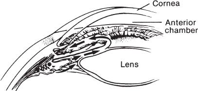
FIGURE 61.1 Normal anterior chamber angle. Aqueous passes from the posterior chamber through the pupil into the anterior chamber and out through the trabecular meshwork.
The iris contains two muscle groups, the radial and circular, whereas the ciliary body contains one group, the ciliary muscle. The radial muscle, which contains α1-receptors, contracts when stimulated by the SNS, resulting in mydriasis. The circular muscle of the iris and the ciliary muscle both contain muscarinic receptors under the control of the PNS. Stimulation of the circular muscle causes contraction and thus miosis. Ciliary muscle contraction reduces the tension on the zonules of Zinn, thereby reducing tension on the lens. Zonule laxity allows the lens to “round up” for accommodation of near vision; however, this also increases the contact between the lens and posterior iris, potentially inhibiting flow of aqueous humor from the posterior to the anterior chamber. Conversely, relaxed tension on the meshwork reduces resistance to outflow at the angle by enhancing the opening from the anterior chamber into the canal of Schlemm.
Elevated IOP in glaucoma most often results from an obstruction to aqueous outflow rather than excessive production (6). Obstruction to outflow from the anterior chamber occurs in both open-angle glaucoma and AACG. The location and mechanism of the resistance to outflow distinguish these two conditions. In open-angle glaucoma, the trabecular meshwork of the anterior chamber angle remains open, but the aqueous humor is unable to reach the canal. In AACG, the peripheral iris contacts the trabecular meshwork and blocks the flow of aqueous into the canal.
Several anatomic features predispose an eye to AACG, and patients may have any combination of risk factors. A shallow anterior chamber results in increased baseline narrowing of the entrance to the angle, with a greater area of contact between the lens and iris. This lens-to-iris apposition impedes the flow of aqueous from the posterior chamber to the anterior chamber through the pupil, produces a forward bowing of the peripheral iris, and effectively closes the angle (Fig. 61.2). Those with a shallow anterior chamber angle, at increased risk for the development of AACG, include the elderly, women, patients of Asian or Inuit descent, and farsighted patients (2,4,5,7). A pharmacologic or physiologic event in a susceptible person typically triggers an attack of acute angle closure by upsetting the equilibrium of aqueous humor dynamics.
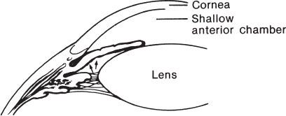
FIGURE 61.2 Angle closure with pupillary block. The angle is closed as the iris is pushed up against the trabecular meshwork.
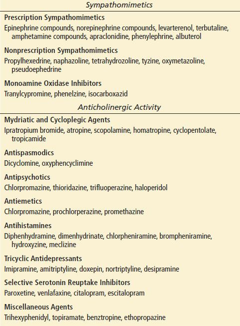
Pupillary dilatation frequently precipitates an attack of acute angle closure. As the pupil dilates, the peripheral iris becomes more flaccid and may be pushed against the trabecular meshwork, blocking aqueous outflow. This may occur when a person is in a dimly lit environment or under emotional stress perhaps due to increased sympathetic tone. Medications that stimulate α1– and/or β1-receptors (sympathomimetics) and those that oppose M3 receptors (anticholinergics) cause pupillary dilatation and may precipitate AACC (Table 61.1) (3,4,8).
TABLE 61.1
Common Pharmacologic Preparations That May Precipitate AACG
CLINICAL PRESENTATION
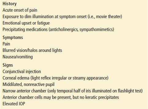
Most patients present with a complaint of sudden onset of severe pain localized to the orbit, or brow or more generally with a severe headache, often associated with nausea and vomiting. Occasionally, the presenting clinical picture is more systemic than ocular, leading to an erroneous or missed diagnosis (9). The physician should consider AACG in patients with headache and high-risk patients with undifferentiated gastrointestinal illness. Blurred vision or the appearance of rainbow-colored halos around lights may occur shortly after the onset of pain (Table 61.2).
TABLE 61.2
Diagnosis of AACG
The patient with AACC has a unilateral red eye with congested episcleral and conjunctival blood vessels, a nonreactive middilated pupil, corneal edema, a shallow anterior chamber, and a high IOP (Fig. 61.3). The IOP is elevated, usually ranging between 60 and 90 mm Hg (IOP <21 mm Hg is considered normal). If the attack has been prolonged, however, the IOP may be lower, due to ciliary body ischemia and decreased aqueous production. Corneal involvement varies with the duration of the attack but is often hazy or steamy appearing from epithelial edema. The anterior chamber appears shallow. Although mild anterior chamber inflammation is common, keratic precipitates (aggregates of white blood cells) on the corneal endothelium are not a typical finding.
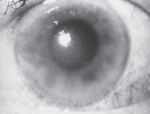
FIGURE 61.3 This eye demonstrates the hallmarks of acute angle-closure glaucoma: perilimbal injection, corneal edema, and a middilated nonreactive pupil.
The course of untreated AACG varies. An attack may damage the optic nerve as well as the corneal endothelium, lens, and retinal ganglion cell layer. In some undiagnosed patients, the attack will continue and, if untreated, can result in permanent blindness within 2 to 3 days. Visual prognosis improves with the initiation of prompt, effective treatment, but any damage to the optic nerve is irreversible.
DIFFERENTIAL DIAGNOSIS
The differential diagnosis of the acutely painful red eye is extensive and is described in detail in Chapter 56. Because headache, nausea, and vomiting may be dominant symptoms in glaucoma, the differential should also include central nervous system and gastrointestinal disorders. Most conditions are readily distinguished by a careful history and physical examination. Other ophthalmic disorders that more closely mimic AACG are the secondary glaucomas: glaucomatocyclitic crisis, neovascular glaucoma, glaucoma secondary to iritis or uveitis, and lens-induced glaucoma.
Glaucomatocyclitic crisis produces recurrent attacks of visual blurring and halos but, in contrast to AACG, pain is not a prominent feature. In addition, the anterior chamber is deep and keratic precipitates develop on the cornea after 2 to 3 days (10).
Neovascular glaucoma presents with a chief complaint of a painful red eye months after vision impairment in the affected eye. It is usually associated with disorders such as, diabetic proliferative retinopathy and retinal vascular occlusion. Retinal hypoxia serves as the underlying stimulus for neovascularization. If the condition progresses, a fibrovascular membrane may obstruct the drainage angle, leading to secondary AACG (4).
Acute iritis with secondary glaucoma presents with ocular pain, photophobia, and blurred vision. The onset is gradual in contrast to AACG. Clinical findings may mimic angle closure, with perilimbal conjunctival injection and corneal edema. However, a miotic pupil, keratic precipitates, and a deep anterior chamber differentiate acute iritis with secondary glaucoma from AACG.
Phacolytic glaucoma presents similarly to AACG, but the anterior chamber depth (ACD) is normal and there are cells in the anterior chamber. Keratic precipitates are also present, and a mature or hypermature cataract can be seen. The cause of phacolytic glaucoma is the release of soluble lens protein into the aqueous, which leads to elevated IOP when it occludes the trabecular meshwork and canal (4,11).
Lens particle glaucoma is associated with spontaneous or traumatic disruption of the lens capsule. As with acute iritis and phacolytic glaucoma, the anterior chamber is of normal depth. In addition, the pupil is often miotic, and lens cortical material is present in the anterior chamber. The particulate lens material reduces outflow in the trabecular meshwork, with resultant increased IOP (4,11).
ED EVALUATION
A careful history is an essential part of the ED evaluation and should include onset of symptoms (sudden, gradual, subsequent to accidental or surgical trauma); history of similar symptoms previously (brief episodes of pain, blurred vision, and/or halos around lights); visual impairment in the affected eye and the unaffected eye; pain in or around the eye; discharge or secretions from the eye; and a detailed medication history.
The ocular examination is described in detail in Chapter 54. The following aspects of the ocular examination deserve special emphasis in relation to AACC and glaucoma. Visual acuity is probably one of the most important evaluations done during any ocular examination. If a distance or near visual acuity cannot be recorded owing to inability to read the chart, the following should be recorded (including the distance at which each is tested): finger counting, and the presence or absence of perception of hand motion and light. Conjunctival injection is often present but is nonspecific. The pattern of injection, however, may be helpful. Perilimbal injection suggests acute iritis or angle closure. Segmental injection characterizes episcleritis; diffuse injection is typical of keratoconjunctivitis. Finally, the presence of purulent discharge suggests an infectious cause. Corneal edema is apparent in many cases of AACG and is due to the rapid rise of IOP. Assessment of the ACD is essential in the evaluation of the painful red eye and in the diagnosis of AACG. In the ED setting, an adequate estimation of the ACD can be accomplished with the oblique flashlight test and slit-lamp examination.
Oblique Flashlight Test
In this test (Fig. 61.4), the examiner shines the light across the eye from the temporal side toward the pupil. In an eye with a normal depth anterior chamber, the entire iris is illuminated. In an eye with a narrow angle or shallow ACD, a shadow is cast on the nasal side of the iris (12).
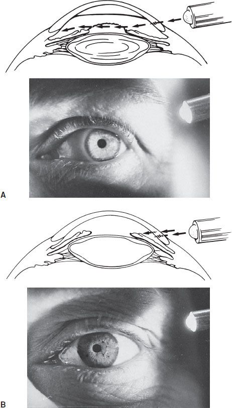
FIGURE 61.4 Flashlight test for wide and narrow angles. A: Wide angle: Entire iris is illuminated. B: Narrow angle: Only temporal half of iris is illuminated. (Photo courtesy of D. R. Anderson, MD.)


