Methylene Blue Injection/Open Joint Evaluation
INDICATIONS
![]() When there is clinical suspicion of communication between a traumatic wound and joint space
When there is clinical suspicion of communication between a traumatic wound and joint space
![]() When intra-articular air within the joint space is seen on radiographs
When intra-articular air within the joint space is seen on radiographs
CONTRAINDICATIONS
![]() Evidence of overlying cellulitis on the site of arthrocentesis
Evidence of overlying cellulitis on the site of arthrocentesis
![]() Patients taking serotonergic psychiatric drugs (risk of serotonin syndrome) and those having glucose-6-phosphate dehydrogenase deficiency (risk of anemia and methemoglobinemia) (FIGURE 49.1)
Patients taking serotonergic psychiatric drugs (risk of serotonin syndrome) and those having glucose-6-phosphate dehydrogenase deficiency (risk of anemia and methemoglobinemia) (FIGURE 49.1)
RISKS/CONSENT ISSUES
![]() Risk of iatrogenic septic arthritis
Risk of iatrogenic septic arthritis
![]() Risk of iatrogenic hemarthrosis
Risk of iatrogenic hemarthrosis
![]() Allergic reaction to local anesthetic
Allergic reaction to local anesthetic
![]() Pain during and after procedure
Pain during and after procedure
LANDMARKS
![]() In most cases approach is via the extensor surface of joints (avoids vessels and nerves)
In most cases approach is via the extensor surface of joints (avoids vessels and nerves)
![]() Specific landmarks should be utilized depending on the joint
Specific landmarks should be utilized depending on the joint
![]() If available, ultrasound can be used to facilitate arthrocentesis
If available, ultrasound can be used to facilitate arthrocentesis
TECHNIQUE
![]() Prepare the Methylene Blue Injection
Prepare the Methylene Blue Injection
![]() Methylene blue usually comes in 1-mL ampoules. There are no exact dilutional guidelines; we recommend diluting 1 mL of methylene blue with 29 mL normal saline in a 30-mL syringe.
Methylene blue usually comes in 1-mL ampoules. There are no exact dilutional guidelines; we recommend diluting 1 mL of methylene blue with 29 mL normal saline in a 30-mL syringe.
![]() Attach an 18- or 20-gauge needle to the syringe
Attach an 18- or 20-gauge needle to the syringe
![]() Prepare the Joint Before Injection
Prepare the Joint Before Injection
![]() Prepare the skin using either a povidone–iodine solution or a chlorhexidine solution and a large sterile drape
Prepare the skin using either a povidone–iodine solution or a chlorhexidine solution and a large sterile drape
![]() Using a small-gauge needle, deposit a small wheal of either 1% or 2% lidocaine for local anesthesia
Using a small-gauge needle, deposit a small wheal of either 1% or 2% lidocaine for local anesthesia
![]() Inject the Joint
Inject the Joint
![]() Enter the joint space using standard arthrocentesis technique
Enter the joint space using standard arthrocentesis technique
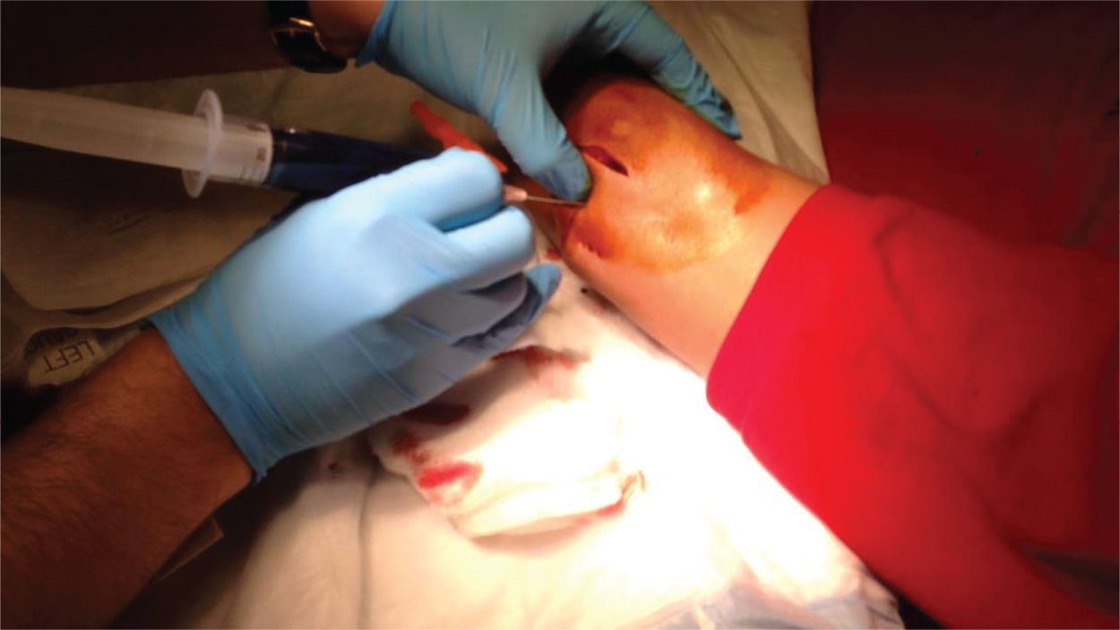
![]() Inject the methylene blue solution into the affected joint space until the joint is fully distended or methylene blue exudes from the wound
Inject the methylene blue solution into the affected joint space until the joint is fully distended or methylene blue exudes from the wound
![]() The amount of solution necessary to fully distend the joint is mostly dependent on the joint in question
The amount of solution necessary to fully distend the joint is mostly dependent on the joint in question
![]() The shoulder can hold ∼30 mL and the knee can hold approximately 60 mL
The shoulder can hold ∼30 mL and the knee can hold approximately 60 mL
![]() Studies support injecting up to the maximum tolerated volume of the joint or until fluid begins to extravasate through the wound
Studies support injecting up to the maximum tolerated volume of the joint or until fluid begins to extravasate through the wound
![]() A positive test is considered if any extravasation of fluid is seen from the wound
A positive test is considered if any extravasation of fluid is seen from the wound

FIGURE 49.1 Arthrocentesis with injection of methylene blue dilution to assess traumatic arthrotomy in a patient with a deep laceration over the elbow. (Courtesy of Felipe Teran, MD.)
Stay updated, free articles. Join our Telegram channel

Full access? Get Clinical Tree


