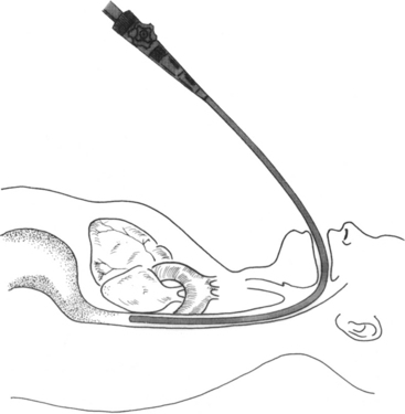
PROCEDURE 79 • Knowledge of cardiovascular anatomy and physiology is necessary. • Knowledge of basic dysrhythmia recognition and treatment of life-threatening dysrhythmias is needed. • Advanced cardiac life support knowledge and skills are necessary. • A topical anesthetic is used in the oropharyngeal area; thus, the patient’s gag reflex may be diminished or absent, putting the patient at risk for aspiration.12,15 • It is essential to understand the institution’s intravenous (IV) conscious sedation guideline. • Sedation can put the patient at risk for respiratory depression.3,5,15 • A fiberoptic probe with an ultrasound transducer is inserted through the mouth and into the esophagus just behind the heart (Fig. 79-1). The transducer located at the tip of the probe sends high-frequency sound waves toward the heart, which return as echoes. The echoes are converted, by computer, into moving images of the heart. The image is displayed on a screen and can be recorded on videotape or compact disk (CD), printed on paper, or sent electronically to a picture archiving communication system (PACS). This test is used to visualize structures of the heart and aorta that may not be seen with a standard transthoracic echocardiogram (TTE) and to clarify structures, that may be otherwise poorly seen. The test may be performed as an outpatient or inpatient procedure or in the operating room.3,16,18 • Various modes of echocardiography are used to examine the heart, blood vessels, valve function, and blood flow. The three techniques are as follows17: • Transesophageal echocardiography (TEE) imaging is more risky than transthoracic imaging because of the insertion of the probe in the esophagus and the need for IV conscious sedation.4,16 • Indications for TEE are as follows:
Transesophageal Echocardiography (Assist)
PREREQUISITE NURSING KNOWLEDGE
 Motion-mode (M-mode) echocardiography: This is a one-dimensional echocardiogram that visualizes time, depth, and intensity. It looks like a tracing instead of a picture of the heart and is used to measure the exact size of the heart chambers.
 Two-dimensional (2-D) echocardiography: This shows the actual shape and motion of the different heart structures. These images represent “slices” of the heart in motion.
 Doppler echocardiography: This assesses the flow of blood through the heart. The signals that represent blood flow are displayed as a series of black-and-white tracings or color images on the screen.
 Evaluation of (pre) clot formation in the heart, especially in the atria and appendages, in patients with an atrial dysrhythmia.3,6,15,18,20
 Evaluation of spontaneous echocardiographic contrast or “smoke” presenting as dynamic echoes within the left atrium and appendage, which resembles swirling smoke in 2-D images. It is manifested by erythrocyte and platelet aggregates in regions of low blood flow; it has a significant correlation with previous embolic events and may serve as a marker for increased risk for embolism.3,6,15,18,20
 TEE before cardioversion is advocated in patients in whom early cardioversion would be clinically beneficial. Patients with atrial fibrillation undergoing electrical cardioversion with short-term anticoagulation therapy have lower hemorrhagic complications. Cardioversion may be performed more safely, after only a short period of anticoagulant therapy, in patients without atrial cavity or appendage thrombus. with TEE. Cardioversion is delayed in patients at high risk with thrombus detected by TEE. Conventional treatment has been to give patients undergoing elective cardioversion therapeutic anticoagulation therapy for 3 weeks before and 4 weeks after cardioversion, to decrease the risk for thromboembolism.11,12,16
 Transient ischemic attack or stroke evaluation to rule out cardiac source of emboli and structure abnormalities (e.g., patent foramen ovale) or other abnormalities not identified before a neurologic event.3,15,16
 Multiple factors may obstruct the penetration of the ultrasound beams from the transthoracic approach. Poor-quality TTE images can be found in patients with obesity, chronic obstructive lung disease, chest wall deformities, multiple chest trauma, and thick surgical chest dressings.3,15
 Assessment of native cardiac valve defects, particularly of the mitral valve.1–3,15
 Assessment of prosthetic cardiac valve function.3,15,16,18
 Assessment of intracardiac foreign bodies, tumors, or masses.3,15,16,18
 Assessment of vegetative endocarditis and abscess.3,8,15,16
 Assessment of congenital heart defects.3,15,16,18
 The superior sensitivity and specificity of TEE for aortic disease, including aneurysm, dissection, atherosclerosis, mobile plaque, congenital aortic disease, pseudoaneurysm, and traumatic aortic disruption, make it the test of choice in many clinical situations.3,15,16,18
 Disease in the ascending and transverse aorta often necessitates a TEE for complete evaluation; however, a short portion of the distal ascending aorta and proximal transverse arch is usually not visible. This portion is a blind area because of the carina passing between the aorta and the esophagus.3,15,16,18
 TEE used in combination with stress test for the evaluation of patients with coronary artery disease. Transesophageal echocardiography–dobutamine stress echocardiography (TEE-DSE) has been reported to be highly accurate for detection of ischemia in patients with suspected coronary artery disease.19
 Transesophageal atrial pacing stress echocardiography (TAPSE) is an efficient alternative to DSE for the detection of coronary artery disease. The heart rate can be rapidly increased, resulting in myocardial ischemia in regions supplied by stenosed coronary arteries. In contrast to TEE-DSE, termination of pacing results in nearly instantaneous restoration of the patient’s intrinsic heart rate.10
 Intraoperative guide to left ventricular function and intracardiac blood flow and evaluation of cardiac surgical repair.13
 Assessment of a donor heart for transplant.18
 Intracardiac shunt evaluation. Right-sided echocardiography saline contrast studies are performed to document an atrial septal defect or a patent foramen ovale and to increase the signal strength of the tricuspid regurgitant jet to allow a more accurate estimate of pulmonary artery pressures.18 Saline contrast for TEE is an IV injection of microbubbles formed by agitating a saline solution. This saline contrast results in a marked increase in echogenicity of the right-sided cardiac chambers.22
 Cardiac assessment in the interventional laboratory during percutaneous interventions, such as transcatheter closure for atrial septal defects and ventricular septal defects.16,18
 Cardiac assessment during interventional procedures, such as balloon mitral valvuoplasty, nonsurgical reduction of the ventricular septum in patients with hypertrophic cardiomyopathy, and transseptal catheterization for placement of a catheter during radiofrequeny ablation of cardiac dysrythmias.16,
![]()
Stay updated, free articles. Join our Telegram channel

Full access? Get Clinical Tree


79: Transesophageal Echocardiography (Assist)

