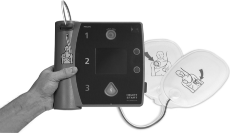
PROCEDURE 39 • Defibrillation is the therapeutic use of an electrical shock that temporarily stops or stuns an irregularly beating heart and allows the spontaneously repolarizing pacemaking cells within the heart to recover and resume more normal electrical activity. Ventricular fibrillation (VF) and ventricular tachycardia (VT) are the only two rhythms recognized as shockable by an automated external defibrillator (AED) (Fig. 39-1). • Time is the major determining factor in the success rates of defibrillation. For every minute defibrillation is delayed, the chance of success decreases by 7% to 10%.1,2,5 When used in conjunction with effective CPR, the decrease in the likelihood of success is more gradual and averages 3% to 4% per minute.1,2,5 • Although defibrillation is the definitive treatment for VF and pulseless VT, the use of the AED is not a stand-alone skill; it is used in conjunction with CPR. CPR should be started as soon as the patient is found to be pulseless and not stopped until the AED has been turned on, the pads have been attached, and the machine is prompting the provider to “stand clear” or “don’t touch the patient.”1,2,5 Immediate postshock CPR starting with compressions has been documented to lead to increased return of spontaneous circulation and increased cerebral survival,4,5 which is why time is not taken to check for a rhythm or pulse after defibrillation. • Ventricular fibrillation depletes the cardiac energy stores of adenosine triphosphate (ATP) more rapidly than a normal rhythm. The longer a heart goes without circulation, the more depleted its energy stores. In a heart with depleted energy stores, defibrillation is more likely to result in asystole because no fuel remains to support spontaneous depolarization or myocardial contraction. Effective CPR can supply the needed oxygen and energy substrates to the heart cells and allow them to return to a perfusing rhythm.4,5 • Three stages of VF are seen in cardiac arrest. The first phase is the electrical phase. During this phase, which is considered the first 4 to 5 minutes of VF, defibrillation is most likely to be effective, and the sooner the shock can be delivered the more likely it is to work. During the next 5 to 10 minutes after VF occurs, the hemodynamic or circulatory phase, a brief period of CPR may “prime the pump” and provide oxygen and energy substrate to the myocardial cells, improving the effectiveness of the defibrillation. If the patient is found during this phase, or if CPR is not ongoing when the defibrillator arrives, effective CPR needs to be administered for approximately 2 minutes, or five cycles of 30 compressions to two ventilations, before the shock. The metabolic phase starts 10 minutes after VF. During this phase, the cardiac cells have experienced global ischemia and energy depletion if no CPR has been initiated. CPR before defibrillation is more likely to be successful and needs to be used in conjunction with advanced cardiac life support (ACLS) therapies.4,5 • The AED is attached to the patient with adhesive electrode pads. Through these pads, the rhythm is analyzed and a shock delivered, if indicated. If the AED recognizes VF or VT, visual and verbal prompts guide the operator to deliver a shock to the patient. The AED, not the operator, makes the decision about whether the rhythm is appropriate for defibrillation. • The chance of the AED shocking inappropriately is minimal.1,5,7 The AED should be applied only to unresponsive, nonbreathing, pulseless patients. To keep artifact interference to a minimum, the patient should not be touched or moved during the analysis time.
Automated External Defibrillation
PREREQUISITE NURSING KNOWLEDGE
Related posts:
![]()
Stay updated, free articles. Join our Telegram channel

Full access? Get Clinical Tree


39: Automated External Defibrillation