Weapons of Mass Destruction


Weapons of Mass Destruction


30-1 Personal Protective Equipment

Anthony Morocco

Personal protective equipment (PPE) is usually classified into four levels based on respiratory and contact protection.1


  • Level A: Self-contained breathing apparatus and completely sealed chemical-resistant suit. This gives the highest level of protection against inhalation or contact with chemical or biologic agents and should be reserved for on-scene rescuers with exposure to vapor respiratory and contact threats.


  • Level B: Self-contained or supplied-air positive-pressure respirator and chemical-resistant suit with gloves and boots. The respiratory protection is equivalent to that of level A, but contact protection is reduced to resist splashes but not vapors. Level B is useful for on-scene rescuers with potential splash and respiratory exposure or oxygen-depleted environments.


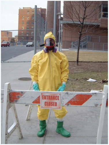
  • Level C: Air-purifying respirator and chemical resistant suit with gloves and boots. Contact protection is equivalent to that of level B, but respiratory protection is decreased. Respirators filter ambient air through cartridges and may be powered to provide positive pressure (greater protection than non-powered respirators). Different cartridges or combination of cartridges are used, depending on the type of agent to be filtered (e.g., high-efficiency particulate air [HEPA] filters). Level C is probably the highest level of protection needed for health care facility workers. It is useful when dealing with patients who are contaminated with chemical or biologic agents.


  • Level D: Standard work clothing and protection, such as hospital scrubs with gown, mask, and gloves (standard universal hospital precautions).


FIGURE 30-1 Level C personal protective equipment with exposure suit, gloves, boots, and mask. (Courtesy of Mark Silverberg, MD.)

The choice of appropriate PPE depends on a number of factors. Higher levels of protection necessitate bulky, heavy, and cumbersome equipment; extensive training and fit-testing for proper use; large expense for equipment and training; lower usable work time due to limited air supply, heat, and exhaustion; difficulty with vision and communication while wearing masks and hoods; and time delays in donning complex suits. For these reasons, the lowest level of protection that is adequate for the potential risk should be employed. In addition, when dealing with patients who are contaminated with radioactive material, health care workers should wear dosimeters.1


REFERENCES

1. Hick JL, Hanfling D, Burstein JL, et al. Protective equipment for health care facility decontamination personnel: regulations, risks, and recommendations. Ann Emerg Med 2003;42:370-380.


30-2 Decontamination

Anthony Morocco

Victims of chemical or biologic weapons may, in some circumstances, require decontamination. Ideally, decontaminations should take place at a site removed from the hospital; however, disaster-level events may result in large numbers of nondecontaminated patients presenting to hospital emergency departments. Therefore, it is vital for hospitals to have adequately trained personnel, appropriate and fit-tested equipment, and a plan for decontamination.

Victims of chemical agents are likely to require more extensive decontamination than those exposed to biologic agents, because chemical agents can be volatile and dermally active. Reaerosolization of biologic particles on victims is possible but is a less immediate risk than chemical exposure.1

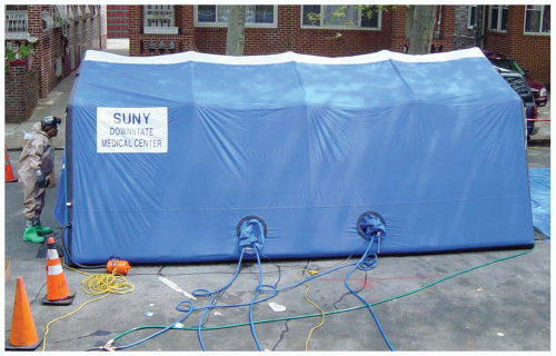
The first step in decontamination at a health care facility is primary triage, which should be performed outside the hospital proper, by personnel wearing appropriate personal protective equipment (PPE). The level of PPE should be tailored to the exposure, if known. For chemical agents, those performing triage and decontamination should wear level C PPE. For biologic contamination, level D is appropriate, with latex gloves, eye protection, and N-95 masks or high-efficiency particulate air (HEPA) filter masks. For unknown events, level C PPE should be used, with a respirator equipped with a combination organic vapor and HEPA cartridge. Patients with no symptoms are triaged to a self-decontamination or waiting area. Ill patients enter the medical decontamination and emergency treatment area. Actual decontamination starts with removal of all clothing, which may actually remove 75% to 90% of the hazardous agent. Attention should be paid to privacy and appropriate packaging of contaminated items and valuables. This is followed by copious rinsing with warm water and, possibly, washing with soap for 1 to 2 minutes. Excessively hot water and harsh scrubbing may enhance chemical absorption and should be avoided.


FIGURE 30-2 Decontamination tent. (Courtesy of Mark Silverberg, MD.)

Hypochlorite solution (0.5%) has been recommended to inactivate both biologic and chemical agents. However, because a contact time of 15 to 20 minutes is necessary to deactivate the chemical agents, damage to injured tissues and eyes may occur, and no evidence of superior efficacy versus soap and water exists. For these reasons, hypochlorite solutions are not recommended as part of routine decontamination procedures. The final steps after washing are to dry the patients, place them in gowns, and place ill patients in the emergency department and others in a predetermined holding area.1


REFERENCES

1. Macintyre AG, Christopher GW, Eitzen E Jr, et al. Weapons of mass destruction events with contaminated casualties: effective planning for health care facilities. JAMA 2000;283:242-249.


30-3 Blast Injury

Michael Greenberg


Clinical Presentation

Explosive and incendiary devices are the weapons of choice for terrorists worldwide. Patients who survive the initial event present for care with a wide variety of injuries affecting multiple organ systems.1,2,3


Pathophysiology

Blast injury (BI) is classified as follows (see Table 30-3). Primary BI is caused by an acute change in ambient pressure emanating from the so-called “blast wave” or “overpressure” wave. Primary BI usually affects the respiratory, gastrointestinal, central nervous, and cardiovascular systems and is rarely seen in survivors, because most people who die from BI die from primary BI. Secondary BI results from objects launched as projectiles secondary to the explosive forces. Tertiary BI is caused when an individual is thrown against a fixed object or the ground, or is injured by crushing forces from a structural collapse. Miscellaneous BI involves burns, chemical exposures, and smoke inhalation resulting from the primary explosion.1,2,3


TABLE 30-3 Mechanisms of Blast Injury


Category


Characteristics


Body Part Affected


Types of Injuries


Primary


Unique to high-order explosives, results from the impact of the overpressurization wave with body surfaces


Gas-filled structures are most susceptible—lungs, gastrointestinal tract, and middle ear


Blast lung (pulmonary barotrauma)


Tympanic membrane rupture and middle ear damage


Abdominal hemorrhage and perforation; globe (eye) rupture; concussion (traumatic brain injury without physical signs of head injury)


Secondary


Results from flying debris and bomb fragments


Any body part may be affected


Penetrating ballistic (fragmentation) or blunt injuries


Eye penetration (can be occult)


Tertiary


Results from individuals’ being thrown by the blast wind


Any body part may be affected


Fracture and traumatic amputation


Closed and open brain injury


Quaternary


All explosion-related injuries, illnesses, or diseases not due to primary, secondary, or tertiary mechanisms


Includes exacerbation or complications of existing conditions


Any body part may be affected


Burns (flash, partial, and full-thickness)


Crush injuries


Closed and open brain injury


Asthma, chronic obstructive pulmonary disease, or other breathing problems from dust, smoke, or toxic fumes


Angina


Hyperglycemia, hypertension


Courtesy of the Centers for Disease Control and Prevention.


Diagnosis

Diagnosis is based on standard advanced trauma life support (ATLS) protocols and the judicious use of imaging and laboratory studies, as dictated by those protocols.


Clinical Complications

Complications include localized and systemic infections (including tetanus), deeply imbedded foreign bodies, limb amputations, post-traumatic stress disorder, and other ill-defined neuropsychiatric sequelae related to BI.


Management

Treatment should comport with standard ATLS protocols, with special emphasis on extensive and meticulous wound débridement as needed.1,2


REFERENCES

1. Argyros GJ. Management of primary blast injury. Toxicology 1997;121:105-115.

2. Covey DC. Blast and fragment injuries of the musculoskeletal system. J Bone Joint Surg Am 2002;84:1221-1234.

3. Mayorga M. The pathology of primary blast overpressure injury. Toxicology 1997;121:17-28.


30-4 Dispersion of Radioactive Materials

Anthony Morocco


Clinical Presentation

Patients may present with traumatic injuries and stress reactions but not acute radiation injury. However, dispersion of radiologic agents may result in internal contamination via inhalation, ingestion, or wound contamination. Direct contact with a radioactive source results in skin burns. The extent and onset of radiologic injury depend on the penetration and dose of radioactivity.1


Pathophysiology

Conventional explosives may be used in a “dirty bomb” to inflict trauma and spread radioactive material over a wide area. A number of different radiologic isotopes are used for industrial, medical, and military applications and therefore may be available to terrorists. Cesium 137, cobalt 60, and iridium 192 are sources within industrial radiographic and radiotherapy equipment. Other radionuclides include tritium, iodine 125, iodine 131, cesium 134, strontium 89, strontium 90, americium, and plutonium.1


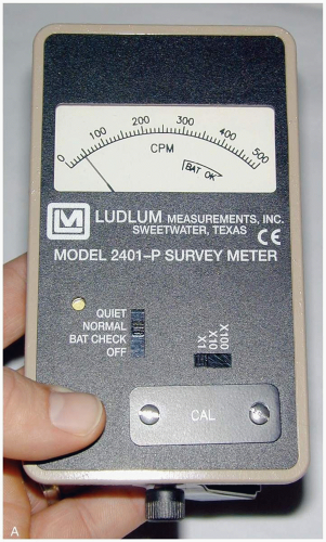
FIGURE 30-4 Radiation survey meter. (Courtesy of Robert Hendrickson, MD.)


TABLE 30-4 Therapy for Specific Isotopes


Radionuclide


Therapy


Tritium


Force fluids


Cesium


Prussian Blue (currently investigational)


Plutonium and transuranics


Chelating agents such as calcium or zinc diethylenetriaminepentaacetate (DTPA) (currently investigational)


Strontium ingestion


Oral aluminum phosphate or barium sulfate


Courtesy of the Department of Homeland Security Working Group on RDD Prep—Medical Preparedness and Response Sub-Group.


Ionizing radiation causes damage to body tissues via free radical formation and direct injury to DNA and other cellular structures.1


Clinical Complications

Radiation doses greater than 6 Gray (Gy) causes bone marrow failure and death without treatment; 10 Gy is the maximum survivable dose with optimal medical care. Malignancy is an important long-term complication. Tumors may develop 5 to 10 years or longer after exposure, whereas leukemia may occur within 2 years.1


Management

The hospital radiation safety officer and the Radiation Emergency Assistance Center/Training Site (REAC/TS, telephone 1-865-576-3131) should be contacted. Victims should be undressed and skin decontaminated. Contaminated wounds should be irrigated, and surgical excision may be necessary for retained particles of long-lived isotopes. Hospital workers are at little risk of exposure to any significant radioactivity. Potassium iodide should be administered within several hours after nuclear detonation or nuclear reactor discharge to prevent thyroid uptake of radioactive iodine. Prussian blue binds cesium and decreases gastrointestinal absorption (see Table 30-4). Chelators (calcium diethylenetriaminepentaacetic acid [DTPA] or zinc DTPA) enhance elimination of americium and plutonium. Strontium absorption is decreased by phosphate administration.1


REFERENCES

1. Mettler FA Jr, Voelz GL. Current concepts: major radiation exposure— what to expect and how to respond. N Engl J Med 2002;346:1554-1561.


30-5 Acute Radiation Syndrome

Michael Greenberg


Clinical Presentation

Patients with acute radiation syndrome (ARS) may be expected to present minutes to hours after exposure to high energy x-irradiation or gamma irradiation. Specific presenting symptoms depend on the total dose of radiation received. Initial symptoms usually include nausea, vomiting, and anorexia.1,2


Pathophysiology

Four clinical phases for ARS have been identified (see Table 30-5B): (1) the prodromal phase, which may range from no symptoms to mild or moderate nausea, vomiting, and diarrhea, depending on degree of exposure, and lasts up to 5 days; (2) the latency phase, which lasts up to 3 weeks and includes dose-dependent hair loss and lymphocytopenia; (3) the phase of overt illness, which includes fatigue, infection, bleeding, thrombocytopenia, hemodynamic instability, coma, and death; and (4) the recovery or demise phase, which occurs after the phase of overt illness.1,2 See Table 30-5A for dermal manifestations.


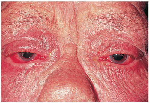
FIGURE 30-5 Erythema, dry scaly skin, and lower ectropion in a patient exposed to local radiation. (From Tasman, with permission.)


TABLE 30-5A Dermal Manifestations of Radiation Dose


Dose (Gy)


Clinical Findings


Time Since Exposure


3


Epilation beginning


14-21 days


6


Erythema


(Transient initially; primary erythema occurs at 14-21 days after exposure)


1-15


Dry desquamation


2-3 wk


20-50


Wet desquamation


2-3 wk


>50


Overt radionecrosis and ulceration


4 wk


From the Department of Homeland Security Working Group on RDD Prep—Medical Preparedness and Response Sub-Group. Gy, Gray.


Diagnosis

Diagnosis is based on history, clinical findings, and results of the complete blood count.


Clinical Complications

Complications are dose-dependent and may include central nervous system dysfunction, cataracts, sepsis, seizures, coma, and death.1,2


Management

Initial treatment may require decontamination efforts followed by meticulous supportive medical care. Because clinical effects are dose-dependent, treatment planning must be individualized, especially if multiple casualties are involved. Cytokine therapy may be helpful in stimulating various bone marrow cell lines that were destroyed or suppressed by radiation exposure.1,2 Bone marrow transplantation may be an option for treatment in some cases.


TABLE 30-5B Acute Radiation Syndromes


Syndrome


Dose


Prodromal Stage


Latent Stage


Manifest Illness Stage


Recovery Stage


Bone marrow (hematopoietic)


0.7-10 Gy (70-1,000 rads)


Mild symptoms may occur as low as 0.3 Gy (30 rads)


Anorexia, nausea and vomiting


Occurs 1 hour to 2 days after exposure


Lasts for minutes to days


Stem cells in bone marrow are dying, although patient may appear and feel well


Lasts 1 to 6 wk


Drop in all blood cell counts for several weeks


Anorexia, fever, malaise


Primary cause of death is infection and hemorrhage


Survival decreases with increasing dose


Most deaths occur within a few months after exposure


In most cases, bone marrow cells begin to repopulate the marrow


There should be full recovery for a large percentage of individuals from a few weeks up to 2 yr after exposure


Death may occur in some individuals at 1.2 Gy (120 rads)


The LD50/60 is about 2.5 to 5 Gy (250 to 500 rads)


Gastrointestinal (GI)


10-100 Gy (1,000-10,000 rads)


Some symptoms may occur as low as 6 Gy (600 rads)


Anorexia, severe nausea, vomiting, cramps and diarrhea


Occurs within a few hours after exposure


Lasts about 2 days


Stem cells in bone marrow and cells lining GI tract are dying, although patient may appear and feel well


Lasts less than 1 wk


Malaise, anorexia, severe diarrhea, fever, dehydration, electrolyte imbalance


Death is caused by infection, dehydration, and electrolyte imbalance


Death occurs within 2 wk after exposure


The LD100 is about 10 Gy (1,000 rads)


Cardiovascular (CV) and central nervous system (CNS)


>50 Gy (5,000 rads)


Some symptoms may occur as low as 20 Gy (2,000 rads)


Extreme nervousness; confusion; severe nausea, vomiting, and watery diarrhea; loss of consciousness; burning sensations of the skin


Occurs within minutes after exposure


Lasts for minutes to hours


Patient may return to partial functionality


May last for hours but often is less


Return of watery diarrhea, convulsions, coma


Begins 5 to 6 hr after exposure


Death within 3 days after exposure


No recovery


LD50/60, dose lethal to 50% to 60% of individuals; LD100, dose lethal to 100% of individuals.


Courtesy of the Centers for Disease Control and Prevention.


Gy, Gray; rads, radiation absorbed dose.


REFERENCES

1. Leikin JB, McFee RB, Walter FG, Edsall K. A primer for nuclear terrorism. Dis Mon 2003;49:485-516.

2. Allen J, Matthews L. Radiation as a weapon of mass destruction. Clin Pediatr Emerg Med 2002;3:248-255.


30-6 Anthrax

Anthony Morocco


Clinical Presentation

Patients with the most serious form of the disease, inhalational anthrax, initially develop nonspecific symptoms such as headache, fever, cough, and chest pain, with progression to dyspnea, hypotension, obtundation, and other signs of severe systemic illness. Cutaneous anthrax begins as a pruritic papule or macule that enlarges, ulcerates, and forms an eschar. Patients with gastrointestinal anthrax may present with ulceration and edema of the mouth and esophagus or with nausea, vomiting, malaise, and bloody diarrhea associated with bowel involvement.1


Pathophysiology

Anthrax is the name for any of the three clinical syndromes caused by Bacillus anthracis, a gram-positive, spore-forming bacterium.1

The bacterium releases three toxins that cause edema, release of inflammatory mediators, hemorrhage, and cell death. Inhalational anthrax occurs after germination of inhaled B. anthracis spores in mediastinal lymph nodes, which may occur up to several weeks after exposure. This form occurs rarely in nature but is likely to develop after exposure to weaponized B. anthracis. Cutaneous anthrax may be transmitted to humans by contact with animals (i.e., “woolsorter’s disease”). The gastrointestinal form occurs after ingestion of improperly cooked, contaminated meat.1

Jun 25, 2016 | Posted by in EMERGENCY MEDICINE | Comments Off on Weapons of Mass Destruction

Full access? Get Clinical Tree

Get Clinical Tree app for offline access