Ventilatory Failure


Ventilatory Failure


▪ PATHOGENESIS OF VENTILATORY FAILURE


Definition

Ventilatory failure is the inability to sustain a sufficient rate of CO2 elimination to maintain a stable pH without mechanical assistance, muscle fatigue, or intolerable dyspnea. Failure to maintain adequate alveolar ventilation usually is recognized by CO2 retention and acidosis. Although a rise in PaCO2 to a level higher than 50 mm Hg has been suggested as definitive, ventilatory failure can occur even when PaCO2 falls to a value lower than its chronic level (which itself may exceed 50 mm Hg). For example, a modest metabolic acidosis may exhaust the limited ventilatory reserve of a patient with quadriplegia, severe airflow obstruction, or acute respiratory distress syndrome (ARDS). In similar fashion, hypocapnic alkalosis may deteriorate to “normal” values for pH and PaCO2 as ventilatory failure develops in a fatiguing asthmatic patient. Conversely, many patients comfortably maintain PaCO2 levels higher than 50 mm Hg on a chronic basis, without satisfying the aforementioned definitions.


Mechanisms of Ventilatory Failure

To maintain effective ventilation, an appropriate signal must first be sent from the brain to the ventilatory muscles. The muscles must then contract with enough force and coordination to generate the fluctuating pleural pressures that drive airflow. The ventilatory power required depends on the difficulty of gas movement and the minute ventilation requirement. Three major mechanisms cause or contribute to ventilatory failure: deficient central drive, ineffective muscular contraction, and excessive workload (Table 25-1). The primary physical signs of ventilatory overstress or fatigue are vigorous use of accessory ventilatory muscles, tachypnea, tachycardia, diaphoresis, and paradoxical motion of the chest or abdomen. The preagonal breathing pattern may be slow, eventually becoming irregular and gasping.


TABLE 25-1 CAUSES OF VENTILATORY FAILURE


Airflow obstruction


Upper airway obstruction


Extrathoracic


Intrathoracic


Functional (OSA)


Lower airway obstruction


Asthma


COPD


Bronchial stenosis (transplant, trauma, tumor)


Muscular weakness


Skeletal muscles


Weakness


Neuromuscular impairment


Quadriplegia


Myopathy


Diaphragm paralysis


Functional


Hyperinflation


Drugs, electrolytes


Ineffective musculature


Thoracic configuration


Chronic


Kyphoscoliosis


Thoracoplasty


Acute


Pneumothorax


Pleural effusion


Flail chest


Hyperinflation


Inadequate ventilatory drive


Intrinsic


Congenital


Chronic loading (obesity, severe airflow obstruction)


Advanced age


Endocrine disturbance


Extrinsic


Drugs/sedatives


Sleep deprivation


Metabolic alkalosis


Nutritional insufficiency


General Principles of Managing Ventilatory Failure

Ventilatory failure is managed by defining its cause, by correcting reversible problems, and by providing mechanical support when required. If the cause of ventilatory failure is not obvious, bedside measurements intended to determine the mechanisms at work are especially important. Ventilatory workload is reflected in the [V with dot above]E and machine pressures needed to deliver the tidal volume (see Chapter 5). Important factors contributing to the minute ventilation requirement include levels of alertness, agitation, pain or discomfort, body size and temperature, pathologic metabolic stress (sepsis, trauma, burns, etc.), ventilatory dead space fraction, nutritional status, and the work of breathing. The difficulty of chest inflation per liter of ventilation is best gauged by the peak dynamic and static (plateau) inflation pressures as well as by the estimated values for resistance, compliance, and auto-PEEP. Neuromuscular function is evaluated by observing the ventilatory pattern, the tidal volume and breathing frequency, and the actions of the respiratory muscles. At the bedside, the appropriateness of ventilatory drive is often best assessed by examining the pH and PaCO2 in relation to breathing effort. (For example, if PaCO2 is high and pH is low, drive may be deficient, muscular reserve may be inadequate, or both; evidence of patient agitation, dyspnea, or distress argues for primacy of the latter.) Integrative indices of demand and capacity, such as the rapid shallow breathing index or the tidal mouth occlusion pressure (P0.1), which is just now coming into clinical use as a quantitative drive index, may be helpful when assessing the continuing need for machine support (see Chapter 10).


Correcting Reversible Factors

The quest to determine the cause for ventilatory failure should be guided by a systematic evaluation of ventilatory drive, [V with dot above]E, the work of breathing, and neuromuscular performance. In passive ventilated patients, resistance and compliance can be measured during constant flow ventilation (see Chapter 5). Therapy to reverse ventilatory failure should be guided by knowledge of the underlying defect and its severity (Table 25-2). Impedance can be improved by relieving airway obstruction (bronchodilation, secretion clearance, placement of a larger endotracheal tube, etc.), by increasing parenchymal compliance (reduction of atelectasis, edema, and inflammation), and by improving chest wall distensibility (drainage of air or fluid from the pleural space, relief of abdominal distention, muscle relaxation, or analgesia). A common problem overlooked in ventilated patients is a closed circuit suction catheter inadvertently left in an advanced position beyond the wye piece. This partial occlusion dramatically narrows the effective caliber of the endotracheal tube and is easily remedied by withdrawing the catheter to its usual position.


TABLE 25-2 REVERSIBLE FACTORS IN VENTILATORY FAILURE


Excessive ventilation requirement


Metabolic acidosis


Increased CO2 generation


Fever


Agitation


Work of breathing


Excessive calories


Increased dead space


Airway apparatus


Hypovolemia


Vascular obstruction


Increased impedance to ventilation


Secretions


Bronchospasm


Airway apparatus


Pleural air or fluid


Abdominal distention


Auto-PEEP


Pulmonary edema


Impaired muscle strength and endurance


Nutritional deficiency


Electrolyte disturbances


POimage, Mg2+, K+


Endocrine disorders


Inadequate cardiac output


Myasthenia gravis/Parkinson disease


Hyperinflation


Drugs (β-blockers, calcium channel blockers)


Impaired ventilatory drive


Drugs (sedatives/analgesics)


Malnutrition


Sleep deprivation


Metabolic alkalosis


Hypothyroidism


Serum chemistries and medication list must be carefully reviewed for potential suppressants of mental status or muscular strength. Neuromuscular efficiency should be optimized by ensuring alertness, maintaining the patient in an appropriate position (usually as upright as possible), relieving pain, and addressing electrolyte disturbances, nutritional deficiencies, and endocrine disorders.
Elevations of ammonia, an endogenous suppressor of consciousness and drive to breathe, should be addressed by reducing correctable sources of its generation—upper GI bleeding, depakote, etc.—by encouraging its gut elimination (lactulose), or by enhancing the metabolism of ammonia to glutamine and hippurate, by using ornithine or benzoate, respectively. Although Addison disease is rare, adrenal insufficiency (absolute or, more commonly, relative) is surprisingly common among critically ill and chronically debilitated patients who undergo major physiologic stress. Measures that improve cardiac output or arterial oxygenation also will improve neuromuscular performance. Treatable neuromuscular disorders (e.g., myasthenia, polymyositis, Parkinson disease) should not be overlooked. Some problems of decreased ventilatory drive are self-limited (e.g., sedative or opiate excess); others improve with nutritional repletion, electrolyte adjustment (metabolic alkalosis), hormone replacement (e.g., hypothyroidism), or recovery of mental status. Very few respond to nonspecific ventilatory stimulants such as progesterone. (Obesity hypoventilation syndrome may be one exception—see following discussion.) Unfortunately, many such problems are refractory to drug manipulation and must be treated by optimizing ventilatory mechanics with the goal of reducing the work of breathing sufficiently to restore compensation.


Mechanical Support

The general principles of intubation, mechanical ventilation with positive pressure, and weaning are presented elsewhere (see Chapters 6 to 7 8 and 10). Noninvasive ventilation offers an attractive option for many patients with mild to moderate disease with rapidly reversible etiologies for ventilatory failure.


▪ SPECIFIC PROBLEMS CAUSING VENTILATORY FAILURE


Airflow Obstruction

Airflow may be obstructed at any level of the tracheobronchial tree. Even in the absence of underlying lung pathology, discrete lesions cause symptomatic airflow obstruction if located at the level of the larynx, trachea, or central bronchi (upper airway obstruction [UAO]). Mediastinal compression because of fibrosis, granuloma, or neoplasia can narrow the trachea or major bronchi. Diffuse diseases of the airways (asthma, chronic bronchitis, emphysema, etc.) usually limit the flow in peripheral air channels (<2 mm in diameter). For certain patients with asthma, however, the primary problem may center on the larynx and upper airway. Airflow obstruction also can occur with such chronic conditions as bronchiectasis, cystic fibrosis, sarcoidosis, and eosinophilic granuloma (histiocytosis). Aspiration, reflux esophagitis, morbid obesity, retained airway secretions, and congestive heart failure (CHF) routinely contribute to airflow obstruction.


Upper Airway Obstruction

Sedentary patients with low ventilation requirements and UAO may remain relatively symptom free until the airway lumen achieves a surprisingly small diameter. Dyspnea then progresses disproportionately to any further decrements in caliber. The complaints of UAO may be difficult to distinguish from those of lower airway disease and may include cardiovascular as well as pulmonary symptoms.


Signs and Symptoms of UAO

The following signs and symptoms are particularly suggestive of UAO (Table 25-3).


  • Inspiratory limitation of airflow.


  • Stridor. This shrill, inspiratory sound is particularly common with extrathoracic obstruction. In an adult, stridor at rest usually indicates a very narrow aperture (diameter < 5 mm).


  • Difficulty clearing the central airway of secretions.


  • Cough of a “brassy” or “bovine” character.


  • Altered voice. Hoarseness may be the only sign of laryngeal tumor or unilateral vocal cord paralysis. (Although not itself responsible, unilateral cord paralysis frequently is associated with processes that do cause obstruction.) Cords paralyzed bilaterally usually meet near the midline, so the voice may be “breathy” or soft but remains audible, despite serious obstruction. Bilateral vocal cord paralysis impairs the ability to generate sound, so the patient must drastically increase airflow for each spoken word. Only short phrases can be spoken before the next breath, and the patient may experience dyspnea when conversing.


  • Marked accentuation of dyspnea and signs of effort by exertion or hyperventilation. The explanation of this nonspecific phenomenon is mechanical. During vigorous inspiratory efforts, negative intratracheal pressures and turbulent inspiratory airflow tend to narrow a variable extrathoracic aperture. Exertion is unusually stressful because obstruction worsens rather than improves during inspiration, as it does in asthma or chronic obstructive pulmonary disease (COPD).


  • Change in breathing symptoms with position changes or neck movement.


  • Failure to respond to conventional bronchodilator therapy and/or steroids.


  • Unexpected ventilatory failure on extubation or precipitous reversal of ventilatory failure by tracheal intubation alone, without ventilatory support.


  • Sudden pulmonary edema.

During asphyxia and severe choking episodes, very forceful inspiratory efforts markedly lower the intrathoracic pressure, increase the cardiac output, and stimulate the release of catecholamines and other stress hormones. The increased loading conditions of the heart, in conjunction with augmented transcapillary filtration pressures, encourage the formation of pulmonary edema.


TABLE 25-3 SIGNS AND SYMPTOMS OF UPPER AIRWAY OBSTRUCTIONa


Inspiratory limitation of airflow


Stridor


Impaired secretion clearance


Brassy or bovine cough


Breathy voice


Disproportionate exercise intolerance


Symptom variation with neck movement


Failure to respond to bronchodilators


Rapid reversal of dyspnea upon intubation


Fulminant episodic pulmonary edema


Frequent panic attacks


a Incidence of these signs will vary with nature, location, and severity of the obstruction.


Diagnostic Tests

The diagnostic workup of UAO may include routine films, computed tomography (CT) or magnetic resonance imaging (MRI) scans of the neck and trachea, and direct visualization by bronchoscopy or laryngoscopy (mirror, direct, or fiberoptic). Reconstructed or 3-dimensional (3D) CT images are often highly informative. Main bronchial obstruction caused by foreign body, tumor, or mediastinal fibrosis may give rise to strikingly asymmetric ventilation and perfusion scans. Similar information may be available through a comparison of full inspiratory with full expiratory chest radiographs. In stable, cooperative patients, pulmonary function tests should include inspiratory/expiratory flow-volume loops, maximal voluntary ventilation, and diffusing capacity as well as routine unforced and forced expiratory spirometry (Table 25-4). Typically, UAO impairs inspiratory flow more than expiratory flow, impairs peak flow and airway resistance disproportionately to FEV1, and responds extraordinarily well to a low-density gas (helium-oxygen) but not well to bronchodilators (unless there is simultaneous bronchospasm). Maximum voluntary ventilation typically is much less than the value predicted from spirometry, whereas vital capacity may be comparatively normal, relative to FEV1.


TABLE 25-4 PULMONARY FUNCTION TESTS SUGGESTING UPPER AIRWAY OBSTRUCTION


Disproportionately reduced peak flow


Maximal midinspiratory flow < maximal


midexpiratory flow


Vital capacity well preserved despite severely


reduced FEV1


Specific airway conductance low despite nearly


normal FEV1


MVVa <30 × FEV1


End-expiratory flows relatively well preserved


DLCO/VAb well preserved


a Maximum voluntary ventilation (L /min).

b DLCO referenced to single-breath lung volume (FRC).


Diffuse airway diseases such as asthma and COPD tend to produce a different pulmonary function test profile. However, asthma can have a significant upper airway component, and occasionally, stridor will be a prominent presenting sign. Often, these patients benefit from anxiolytics or psychotropic drugs as well as bronchodilators and steroids. Unlike the diffuse obstructive diseases, which alter lung volume, distribution of air-flow, and diffusing capacity, UAO tends to leave the parenchyma unaffected. Diffusing capacity is relatively well preserved.

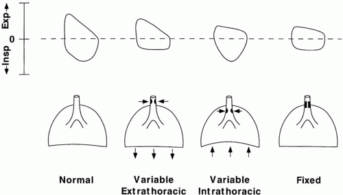
The flow-volume loop contour depends on (a) the fixed or variable nature of the obstruction and (b) the intrathoracic or extrathoracic location (Fig. 25-1). A fixed lesion inside or outside the thorax blunts the maximal inspiration and maximal expiration to a similar degree,
giving a “squared off” loop contour. A variable extrathoracic lesion, surrounded by atmospheric pressure, retracts inward when subjected to negative inspiratory airway pressure but dilates when exposed to positive airway pressure. Conversely, a variable intrathoracic lesion, surrounded by a pleural pressure more negative than airway pressure, dilates on inhalation. On exhalation, the lesion is pushed inward to critically narrow the airway. Unilateral obstruction of a main bronchus may not generate such characteristic curves.


FIGURE 25-1 Flow-volume loops in UAO. Maximal rate of inspiratory airflow is disproportionately curtailed as negative tracheal pressure accentuates resistance through a variable extrathoracic lesion. In similar fashion, the positive pleural pressures generated during forced exhalation selectively limit the airflow across a variable intrathoracic lesion. A fixed lesion at either site limits the maximum flows in both phases.


Management of UAO

The basic principles of managing UAO can be summarized as follows: Patients with symptoms at rest should be kept under continual surveillance and well monitored until the acute crisis resolves. Although certainly indicated, pulse oximetry may give a false sense of security, as O2 saturation may remain within broad normal limits until the brink of total airway obstruction, physical exhaustion, or full respiratory arrest is reached. Postextubation glottic edema and laryngeal swelling resulting from injury usually peak within 12 to 24 h and then recede over the following 48 to 96 h. Racemic epinephrine aerosols may help reduce glottic edema as they cause topical vasoconstriction and bronchodilate the lower airway to reduce the vigor of breathing efforts. For spontaneously breathing patients not already receiving ventilatory support, continous positive airway pressure (CPAP) or BiPAP delivered by mask is often helpful. Upright positioning is favored. For unusually labile or otherwise precarious patients, intubation and tracheostomy kits, as well as a 14-gauge needle (for cricothyroid puncture), should be at the bedside for emergent use. In an emergency, oxygen can be insufflated via the needle until an airway is secured (see Chapter 6). Relief of bronchospasm is particularly important in the setting of a UAO. Relief of lower (small) airway obstruction reduces the intrapleural pressure swings and the severity of upper airway (particularly extrathoracic) obstruction. If there is inflammatory obstruction, tactile stimulation of the involved region must be avoided, and steroids may be helpful. Heliox may also be a reasonable option, especially for those who cannot tolerate or do not respond well to pressurized masks and whose oxygen exchange is well preserved. The patient should be kept calm but alert in a head-up posture. Endotracheal intubation or tracheostomy may be needed if ventilatory failure ensues or secretions cannot be cleared. These procedures should be attempted only by experienced personnel. For otherwise stable patients in whom the airway is “high risk” or known to be difficult to intubate, consideration should be given to conducting intubation and/or extubation in an operating environment where the full range of instruments and supporting measures is available.

Only gold members can continue reading. Log In or Register to continue

Stay updated, free articles. Join our Telegram channel

Jul 17, 2016 | Posted by in CRITICAL CARE | Comments Off on Ventilatory Failure

Full access? Get Clinical Tree

Get Clinical Tree app for offline access