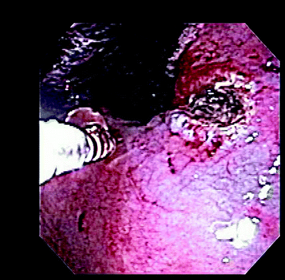
Fig. 8.1
UGI bleeding from a Dielaufoy’s lesion. © Dale Dangleben, MD
Complications
Estimated to occur with a frequency of 0.03%, esophageal perforation is the most troublesome complication of endoscopy. This iatrogenic injury usually occurs in the cervical esophagus (within Killian’s triangle). Hemorrhage is the leading cause of death associated with peptic ulcer disease. Mortality rates for emergent operations for duodenal ulcer tend to be lower than the gastric counterpart since patients are younger and have fewer comorbid conditions. Obstruction and perforation are the other two common complications.
Management
Resuscitation with isotonic fluid is of the first priority in the acutely bleeding patient. In the setting of hemorrhagic shock, definitive airway management may be necessary secondary to diminished mental status and potential aspiration from ongoing emesis. Reversal of possible existing coagulopathy must follow. Nasogastric tube decompression should be performed. Lavage may prevent further bleeding; however, there is no clear evidence supporting the use of room temperature versus iced fluid. Laboratory studies including a type and cross and coagulation parameters along with a serologic test for Helicobacter pylori should be ordered. Continuous monitoring in the intensive care unit is usually required. Early endoscopy continues to be controversial, but undoubtedly helps determine the underlying cause. Intubation before endoscopy may be needed. The therapeutic options during EGD depend on findings. Various methods are available including injection with dilute epinephrine, heater probe or argon plasma coagulation as well as sclerosis and clip application (Fig. 8.2) [1].

Fig. 8.2
Cauterizing a bleeding ulcer. © Dale Dangleben, MD
For variceal bleeding, intravenous infusion of vasopressin (0.4–0.6 units/min) and octreotide (50 μg IV bolus, then 25–50 μg/h IV) is utilized. Endoscopic band ligation and sclerotherapy are options for therapy with repeated endoscopic evaluations 1–2 weeks until varices are obliterated. Non-selective beta blockers are recommended in patients with portal hypertension to prevent recurrence, but have no role in the acutely bleeding patient [2].
Stay updated, free articles. Join our Telegram channel

Full access? Get Clinical Tree


