Fig. 29.1
(a) Dissection of muscles in the buttock. (b) Dissection to the popliteal nerve for open technique nerve placement
29.2 Technical Overview
Ultrasound guidance technology can be used for many peripheral nerve targets, including the occipital nerve, ilioinguinal nerve, intercostal nerve, axillary nerve, median nerve, ulnar nerve, sciatic nerve, posterior tibial nerve, common peroneal nerve, and saphenous nerve. The basic principal is the same: First, the nerve location is identified under ultrasound guidance and then a peripheral electrode is placed in closed proximity. It has most often been observed that best stimulation is achieved by placing the electrode perpendicular to the course of the nerve. Initially, a three and one half–inch 22G spinal needle is used to visualize the path, and then a 14G needle should be placed in the same direction under ultrasound guidance. The 22G needle is used as a safety precaution in the event of invasion of the nerve or a blood vessel.
29.3 Technique Examples
Ultrasound guidance is especially helpful for initial placement of the PNS lead, as illustrated by the following examples.
29.3.1 Occipital Nerve Stimulation
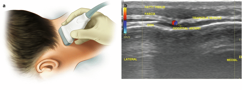
The patient is placed in the prone position. The ultrasound probe is placed just lateral to the occipital protuberance bilaterally. The probe is gradually moved laterally to visualize the pulsation of the greater and lesser occipital artery. The nerve is in close proximity to artery pulsation and on the medial aspect in most patients. In the plane approach, the lead is placed perpendicular to the course of the occipital nerve (Fig. 29.2).


Fig. 29.2
(a) Out of plane position of transducer to locate the occipital nerve. (b) Highlighted US image of greater occipital nerve, and the surrounding anatomy
29.3.2 Ilioinguinal Nerve Stimulation
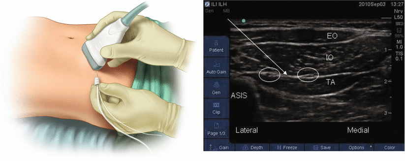
The patient is placed in the supine position. The ultrasound probe is placed 2 cm medial and 2 cm inferior to the anterior superior iliac spine. Under ultrasound guidance, the plane between the internal oblique muscle and the transverse abdominis muscle is visualized and its depth is gauged and marked. At the same depth, the lead is passed under ultrasound guidance in the same plane and perpendicular to the inguinal ligament (Fig. 29.3).


Fig. 29.3


Transducer approach and US image of ilioinguinal anatomy
Stay updated, free articles. Join our Telegram channel

Full access? Get Clinical Tree


