7 Treating muscles, fascia and myofascial trigger points
Myofascial pain is defined as pain arising from muscles or related fascia and comes from hyperirritable areas of muscle, ligaments and fascia known as myofascial trigger points (Bennett 2007). There are approximately 400 muscles forming the largest organ in the body amounting to approximately 40% of the body weight and there is no single medical specialty that is solely responsible for the study of their diagnosis and function. Myofascial trigger points have been associated with low back pain, neck pain, tension headaches, temporomandibular joint pain, forearm and hand pain and pelvic and urogenital pain syndromes in 44 million Americans and so a clinical practitioner in any specialty is likely to see patients with myofascial pain caused by trigger points (Borg-Stein 2006, Simons 1983, Fernandez-de-las-Penas 2006, 2007, Ardic 2006, Hwang 2005, Dogweiler-Wiygul 2004).
Characteristics of myofascial trigger points
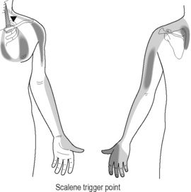
Active myofascial trigger points cause pain at rest, restrict muscle range of motion and have characteristic myotomal pain referral patterns that do not follow dermatomal nerve root patterns or scleratomal patterns emanating from joint structures. The original referral patterns were identified in the 1930s by injecting hypertonic saline into muscles (Kelgren 1938) and pain patterns for over 100 muscles have been documented in detail in the two-volume text, Myofascial pain and dysfunction: the trigger point manual (Travell 1983, 1992). Myofascial trigger points are found by gentle palpation across the direction of the muscle fiber to identify an indurated taut band of muscle and then specific palpation within the taut band to locate a painful nodule that feels like a hardened grain of rice or lentil. Firm pressure on the small nodule may cause the muscle to twitch and may recreate the patient’s pain complaint in the myotomal referral area. Latent myofascial trigger points have taut bands that are tender to touch and restrict range of motion but do not cause spontaneous referred pain.
Calcium release
Myofascial trigger points are thought to arise from focal injury to muscle fibers caused by trauma or overuse. Biopsies of myofascial trigger points reveal a cluster of numerous microscopic foci of sarcomere “contraction knots” that are scattered throughout the tender nodule (Simons 2001, Gerwin 2004). These contraction knots are thought to be caused by calcium release from the sarcoplasmic reticulum and are maintained by an “energy crisis” in the now hypermetabolic muscle once the constant contraction is initiated. Muscle contraction requires the energy of four ATP; muscle relaxation requires two ATP (Adenosine triphosphate, ATP, is the chemical energy that fuels all physical processes; Guyton 1996). Once facilitated, the motor endplates release increased amounts of acetylcholine to maintain the contraction, perpetuating the contraction knots and forming a self perpetuating cycle of activation, energy depletion and local metabolic stress (Mense 2003). Calcium release from the sarcoplasmic reticulum becomes relevant for FSM treatment of trigger points because the frequency thought to “remove calcium ions” is one that softens the taut band and usually eliminates the trigger points.
Nerve sensitization
Persistence of the trigger point leads to neuroplastic changes at the level of the dorsal horn in the spinal cord, leading to central pain sensitization and expansion of the pain beyond its original boundaries into the referred pain area (Arendt-Nielsen 2003). The central neuroplastic changes account for the characteristic trigger point referral patterns. Neuropathic pain sensitization at the level of the nerve root and the spinal cord accounts for the pain intensity seen during stimulation of the trigger point that often appears disproportionate to the stimulus (Curatolo 2006). The twitch response that occurs when the muscle is stimulated is a spinal reflex that can be abolished by transection of the spinal nerve that innervates the trigger point (Hong 1994, 1996).
The local biochemical milieu in an active trigger point is different from that of normal muscle fibers or latent trigger points. A microdialysis needle was used to take constant stream samples of the biochemical environment within an active trigger point before, during and after a twitch response and compared it to normal muscle and latent trigger points. Active trigger points show significantly elevated levels of the inflammatory peptides TNF-α, Interleukin-1(IL-1), calcitonin-gene-related-peptide (CGRP), substance P, bradykinin, serotonin, and norepinephrine (Shah 2005). Early biopsies of trigger points showed mast cells degranulating releasing histamine into the area around the trigger point (Simons 1983). The neural component of trigger point pain and perpetuation is relevant to FSM treatment because the most effective treatment protocols have evolved to include treating “inflammation in the nerve and the spinal cord” first with the FSM treatment protocols known to reduce inflammatory cytokines (McMakin 2005).
Treating myofascial trigger points
There is no form of drug therapy that alleviates myofascial trigger point pain or muscle dysfunction. Trigger point injections with saline and 1% lidocaine or procaine or dry needling are considered to be the most effective therapy but require a skilled well trained therapist to precisely localize the active trigger point by identifying a local twitch response in the taut band. Studies have shown problems with localizing the taut band and the active trigger point (inter-rater reliability) between therapists depending on their skill and training (Gerwin 1997, Hsieh 2000, Sciotti 2001). Needling or injection of single active trigger points limits effectiveness in muscular areas populated by multiple active, latent and satellite trigger points. Full length stretching of the muscle while using an ethyl chloride vapocoolant spray, called “spray and stretch”, disrupts the focal contractions and stops the prolonged ATP consumption that perpetuates the contraction knots. Not all muscles are suitable for this intervention and environmental considerations have reduced its use in recent years. Postural and ergonomic corrections to modify factors that perpetuate trigger points are critical to successful management.
Diagnosing myofascial pain
Any patient who presents with a chronic pain complaint should have a focused neuromuscular evaluation that includes evaluation of reflexes and sensation and a palpatory evaluation to check for taut bands and myofascial trigger points. There are wall charts and diagrams in text books that illustrate the referred pain patterns for specific muscles (Travell 1983, 1992, Niel-Asher 2008). The diagrams give guidance as to what muscles are likely to be a source of the referred pain in a given area. The practitioner should match the patient’s area of complaint with the pain patterns in the diagram and then check the muscles that refer pain to that area. Palpation of a taut band and the small painful nodule that is the trigger point, pressure on the nodule that reproduces the patient’s pain and restricted range of motion due to muscle tightness are all diagnostic of an active myofascial trigger point.
Treating myofascial trigger points with FSM
FSM was first used to successfully treat myofascial trigger points in 1996 and the first two articles published were collected case reports showing successful pain resolution in trigger points in the head, neck and face pain and in low back pain (McMakin 1998, 2004). FSM provides microamperage current known to increase ATP production by 500% in rat skin (Cheng 1982). The current alone would address the energy crisis that perpetuates the contracture knots allowing the knots to release by increasing ATP.
The frequencies used to “reduce inflammation in the nerve” have been shown to decrease inflammatory cytokines including IL-1, CGRP, substance P and serotonin – demonstrated by Shah to be increased in the active trigger point milieu (McMakin 2005, Shah 2005). FSM has been shown to down regulate spinal cord activation and reduce central sensitization in the treatment of fibromyalgia associated with spine trauma and could reasonably be assumed to perform the same function to reverse the central neuroplastic changes seen in myofascial pain. The observed effects of muscle softening, relaxation of the taut band and resolution of the trigger point occur as a response to specific frequencies meant to reduce inflammation in the nerve and reduce calcium ion deposits in the fascia. It is reassuring that these frequencies coincide with the pathologies now known to be associated with myofascial trigger points.
Treating chronic myofascial pain and trigger points

• The patient must be hydrated to benefit from microcurrent treatment.
• Hydrated means 1 to 2 quarts of water consumed in the 2 to 4 hours preceding treatment
• Athletes and patients with more muscle mass seem to need more water than the average patient.
• The elderly tend to be chronically dehydrated and may need to hydrate for several days prior to treatment in addition to the water consumed on the day of treatment
• DO NOT accept the statement, “I drink lots of water”
• ASK “How much water, and in what form, did you drink today before you came in?”
• Coffee, caffeinated tea, carbonated cola beverages do not count as water.
Channel B: tissue frequencies
Neuropathic component
 Dermatomal nerve roots become inflamed and sensitized from constant input by the contracting myofascial tissue or from a nearby disc injury. The disc nucleus contains very concentrated levels of the inflammatory substance phospholipase A2 (PLA2). When the annulus is damaged by trauma or postural strain it allows diffusion of small amounts of PLA2 to the nerve. This concentration is sufficient to cause nerve inflammation and muscle hypertonicity but insufficient to cause a classic dermatomal neuropathy.
Myofascial component
 The fascia is the thin connective tissue covering surrounding the muscles and all soft tissues. The fascia becomes inflamed, calcified and fibrosed during the degenerative process.
• Artery and Elastic Tissue in the Muscle Belly: __ /62
 62 is the frequency used for the artery and the elastic tissue in the arterial walls. The muscle belly responds to this frequency either because it is full of small arteries or because the elastic tissue in the muscle belly is somehow related to the artery wall.
 Fine neural fibers travel between layers of fascia in a fascia–nerve–fascia sandwich to innervate the muscles. Constant contractures and local metabolic dysfunction create inflammation at the site of the trigger point. Inflammation leads to calcium influx and fibrosis. Fibrosis between the nerve and fascia restricts movement, creates neuropathic pain and muscle activation when the nerve is stretched as the fascia moves. Calcium ions flow into the nerve when it is inflamed and change the firing threshold.
 This frequency appears to influence the connective tissue that creates the matrix for the muscles and fascia.
Joint component – facet joints, discs, and peripheral joints
 The periosteum lines the outside of the bone, interweaves with tendinous and ligamentous attachments and is very pain sensitive. Inflammation and calcification in the periosteum are the most common chronic pain generators in facet and peripheral joint pain.
 The joint capsule attaches to the periosteum, surrounds the joint and becomes fibrosed, calcified, scarred and inflamed when damaged by trauma or chronic mechanical stress.
 Cartilage lines the facet and peripheral joint surface and becomes damaged, degenerated, calcified and inflamed when traumatized by acute or chronic compression of the joint surface (see Chapter 6: Treating Facet Joint Pain).
 The disc annulus wraps around and contains the nucleus and is very well innervated and pain sensitive. It is the most common pain generator in chronic discogenic pain and in mild acute disc injuries that would create myofascial trigger points (see Chapter 4: Treating Discogenic Pain).
 The gel like disc nucleus fills the center of the disc and absorbs water to become a cushion for the vertebral bodies in the spine. It is very high in PLA2 and very inflammatory.
• Bursa or tendon sheath: ___ / 195
 The major tendons in the peripheral joints are cushioned by bursas that lie between the tendons and between the tendons and the periosteum. Bursas become inflamed and calcified by overuse and repetitive stresses.


The 58/’s will increase range of motion but do not change pain.

Caution: 58 / 00, 01, 02, 32
• Do not use these frequencies on injuries newer than 5 to 6 weeks old.
• Newly injured tissue must form scar tissue in order to repair itself.
• Removing the scar tissue seems to undo the healing by weeks in a new injury.
• The 58/’s can be used very briefly (15 seconds) to modify scar tissue as it is forming after the first four weeks.
• Never use this combination before the injury is 4 weeks old

Treat the nerve
Nerve protocol
40 / 396
• Reduce Inflammation in the nerve.
• Polarize current positive +.
• Treatment time: 5 minutes or as long as positive response occurs.
• The protocol to reduce inflammation in the nerve usually begins softening the muscles between the two contacts within one minute. At the end of 5 minutes the softening should have maximized and achieved whatever reduction in neural inflammation or sensitization that can be accomplished.
40 / 10
• Reduce inflammation in the spinal cord.
• Polarize current positive +.
• Treatment time: 2 minutes or as long as positive response occurs.
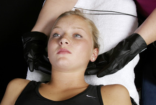
• The protocol to reduce inflammation in the cord is especially helpful in treating muscles in the cervical spine and shoulder that are especially tight bilaterally and seems to address spinal cord sensitization and upregulation. If this frequency combination is going to produce additional softening of the muscles it will do so within the first 2 to 3 minutes. If no change in muscle tone or texture becomes apparent in that time, change frequency to the next in the protocol. If the muscle softens in response to this frequency, the frequency combination can be used until no further softening is perceived which may take up to 5 to 10 minutes depending on the patient.
• Predicted Response: The frequencies for inflammation in the nerve and cord cause muscle relaxation in more than 80% of patients treated. As this response begins, use the manual techniques described below and wait for the tissue to stop softening before changing to the muscle protocol.

Treatment application
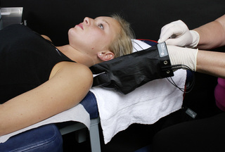
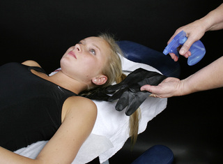
Lead placement: manual therapy
• The positive leads from channel A and channel B are placed in one glove.
• The negative leads from channel A and channel B are placed in the other glove.
• The two gloves will always be on opposite sides of the body part being treated during manual treatment of trigger points and myofascial tissue. The required interferential field forms in the space between the two gloves which should include the tissue being treated. The polarized positive current used with 40 / 396, 10 relaxes the tissue even if the contacts are not set up proximal distal.
• Positive leads: The positive leads are wrapped in or attached to a warm wet contact, such as a hand towel or long graphite electrode that is wrapped around the neck or placed along the spine where the nerve roots exit that innervate the involved muscle group.
• Negative leads: The negative leads are wrapped in or attached to a warm wet fabric contact or long graphite electrode that wraps around the end of the nerve root at the distal end of the muscle group to be treated.
A diagram for the placement would look like this:
| Positive Electrode Channel A | Positive Electrode Channel B |
| Area to be Treated | |
| Negative Electrode Channel B | Negative Electrode Channel A |
Current level
• Average patients require 100–300μamps. Use lower current levels, 20–60μamps, for very small or debilitated chronically ill patients. Use higher current levels, 300–500μamps, for larger or very muscular patients. In general, higher current levels reduce pain and create softening more quickly. Do not use more than 500μamps as animal studies suggest that current levels above 500μamps reduce ATP production (Cheng 1982).
• Note: Current levels over 150μamps make it difficult to use the graphite gloves directly on the skin because they dry out quickly and the current becomes uncomfortable easily. It becomes cumbersome and tedious to continually moisten the graphite gloves. If higher current levels are required due to the patient’s size using wet towel contacts is recommended.
Waveslope
• Use a moderate to sharp waveslope with a ramped square wave pulse.
• The waveslope refers to the rate of increase of current in the ramped square wave as it rises from zero up to the treatment current level every 2.5 seconds on the Precision Microcurrent and the automated family of FSM units. Other microcurrent instruments may have slightly different duty cycles and the waveslope may be different but any unit which provides current flow with a square wave pulse should produce the desired effect. A sharp waveslope has a very steep leading edge on the square wave indicating a very sharp increase in current. A gentle waveslope has a very gradual leading edge on the waveform indicating a gradual increase in current.
• Use a moderate to sharp waveslope for chronic pain. Use a gentle waveslope for new injuries. A sharp waveslope is irritating in new injuries.
• Different microcurrent devices may provide different wave shapes and waveslopes but the reader may find them to be equivalent.