Fig. 23.1
Optimal patient positioning for tracheostomy

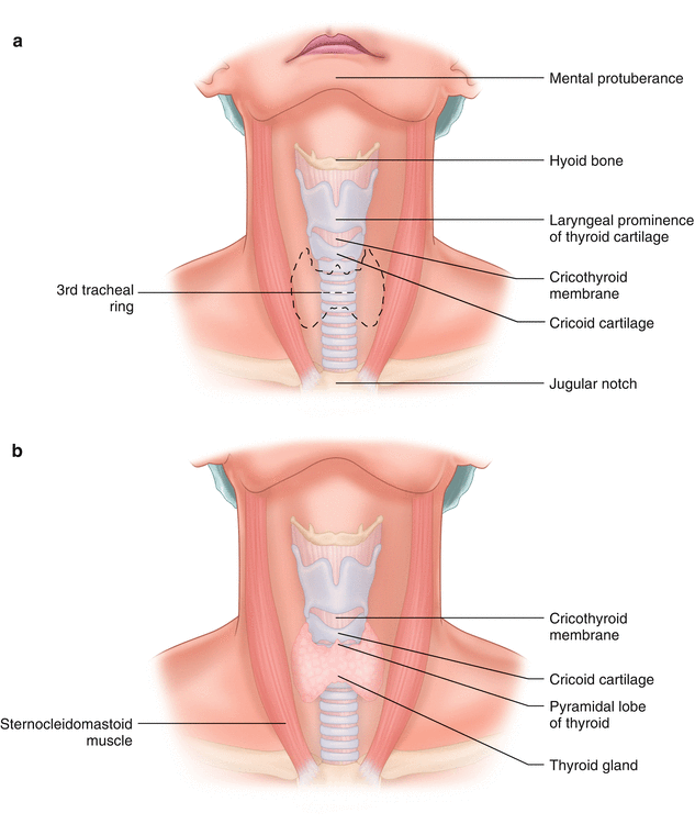
Fig. 23.2
Important neck anatomy for tracheostomy
Tracheostomy
Operative Technique
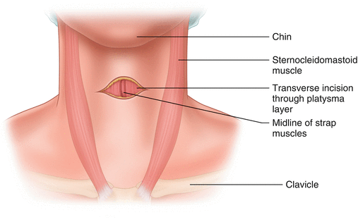
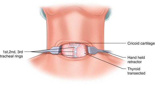
A transverse incision is made two finger breadths above the sternal notch. This transverse incision is taken down through the platysma and then all work subsequently is in the midline between the strap muscles (Fig. 23.3). The midline trachea is identified. The optimal placement is through the third tracheal ring, although this may be difficult in some patients based upon the length of their neck and their anatomy. Typically the isthmus of the thyroid is overlying the second and third tracheal ring. Division of the isthmus allows visualization of the third tracheal ring and mobilization of the thyroid gland off the trachea laterally. The isthmus is transected with cautery and then suture ligated with a braided suture on a robust needle (an MO-6 needle) (Fig. 23.4). This suture is then used for retraction of the soft tissues to gain better access to the trachea.


Fig. 23.3
Transverse incision two fingers above clavicular notch

Fig. 23.4
Isthmus of the thyroid often must be divided
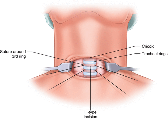
Stay sutures are placed around the third ring if possible, or the second if necessary. At this point in the procedure there is the risk of puncturing the endotracheal tube balloon. The risk of balloon perforation may be diminished by having anesthesia personnel untape the endotracheal tube and deliver the endotracheal tube deeper into the trachea so that the balloon will assuredly be below the level of the operative exposure. The stay suture should be placed with the same robust needle around the third tracheal ring as laterally as possible. The sutures that were used to retract the isthmus of the thyroid are then cut. An H-type incision is made through the third tracheal ring (Fig. 23.5). A tracheostomy spreader is then carefully used. The endotracheal tube is visualized and then slowly withdrawn. Once the endotracheal tube is pulled back to just proximal to the opening in the trachea, the tracheostomy tube is inserted. Some surgeons prefer to deliver the tube into the trachea when the angle is facing the patient’s head and then rotate it 180° once it is in the trachea and deliver it down into the trachea (Fig. 23.6). The tracheostomy tube is then rotated into final position. When placing the tracheostomy tube, the only retraction necessary is the two stay sutures in the lateral trachea.


