Toxicology


Toxicology


Nicholas M. Mohr

Devin P. Sherman

Steven L. Brody


More than 2.5 million cases of accidental or intentional poisoning were recorded by U.S. poison control centers in 2008, resulting in 1315 deaths and over 93,000 intensive care unit admissions. Among fatalities, 91% were 20 years of age or older. Deaths were most common following ingestion of sedatives/hypnotics/antipsychotics (14.8%), opioids (12.7%), acetaminophen alone or in combination (12.7%), antidepressants (7.8%), cardiovascular drugs (7.5%), ethanol (6.7%), and stimulant street drugs (5.8%). Although much of the initial management is provided in the emergency department (ED), critical care physicians may subsequently manage the poisoned patient.


KEY STEPS IN THE MANAGEMENT OF THE POISONED PATIENT

Algorithm 33.1 presents an outline for managing the poisoned patient.


  • Before all else, address the “ABCs” (airway, breathing, and circulation):


    • Intubate and mechanically ventilate for airway protection or respiratory failure


    • Obtain intravenous access and give crystalloid for hypotension. Obtain electrocardiogram (EKG) and routine labs. Obtain extra tubes of blood for serum drug and toxin levels.


  • Treat potentially reversible causes of altered mental status or coma with:


    • Rapid glucose assessment and treatment, if indicated (prior to intubation if possible)


    • Thiamine 100 mg IV


    • Naloxone 0.4 to 2 mg IV or IM if possible opioid toxicity


  • Assess for and treat toxidrome and give specific antidotes when indicated (see Tables 33.1 and 33.2). Call the regional Poison Control Center at 1-800-222-1222 for management advice.


  • Block absorption of toxins when appropriate (see “Gastric Decontamination”).


  • Enhance elimination of toxins when appropriate (see “Enhancing Drug Elimination”).


EMERGENCY EVALUATION

The initial assessment of the poisoned patient should begin with an assessment of the ABCs: airway, breathing, and circulation. Respiratory depression, loss of airway protective reflexes, and aspiration are common consequences of ingestion. Awake patients may need close monitoring for delayed drug effects, and lethargic or obtunded patients,

or those with recurrent seizures may require immediate endotracheal intubation. When in doubt, the airway should be secured by intubation. Care should be taken to clear the airway of secretions or obstruction.


ALGORITHM 33.1 Key Steps in the Initial Management of the Poisoned Patient


TABLE 33.1 Clinical Toxidromes


Possible features


Offending agents


Sympathomimetic


Hypertension, tachycardia, tachypnea, hyperthermia, mydriasis, agitation, hallucinations, diaphoresis


Cocaine Amphetamines Ephedrine Pseudoephedrine Theophylline Caffeine


Anticholinergic “Hot as a hare, dry as a bone, red as a beet, mad as a hatter.”


Hypertension, tachycardia, tachypnea, hyperthermia, mydriasis, agitation, delirium, hallucinations, dry skin, dry mouth, ileus, urinary retention


Tricyclic antidepressants Antihistamines Atropine Phenothiazines Scopolamine Belladonna alkaloids


Cholinergic “SLUDGE”


Salivation, lacrimation, urination, diarrhea, gastrointestinal distress, emesis; also bradycardia, miosis, confusion, coma, bronchoconstriction


Organophosphates Physostigmine Pyridostigmine Edrophonium


Opioid


Hypotension, bradycardia, hypopnea, bradypnea, hypothermia, miosis, CNS depression/coma, decreased bowel sounds, pulmonary edema


Heroin Oxycodone Morphine Meperidine Fentanyl Codeine Methadone


Sedative-hypnotic


Hypotension, bradycardia, hypopnea, bradypnea, CNS depression, coma


Benzodiazepines Barbiturates Alcohols


Extrapyramidal


Rigidity, torticollis, opisthotonos, trismus, oculogyric crisis, dysphoria


Prochlorperazine Haloperidol Chlorpromazine Other antipsychotics


Ventilatory failure in the poisoned patient may be a consequence of respiratory depression (e.g., from sedatives) or muscle paralysis/weakness (e.g., from botulism). Arterial blood gas (ABG) measurement will reveal an elevated PaCO2. Somnolence or obtundation in the setting of a rising PaCO2 is an indication for intubation and assisted ventilation. Bronchospasm may be observed in cases of inhalational injury and other toxins, and bronchodilators may be useful.


TABLE 33.2 Selected Causes of an Elevated Anion Gap


Toxic ingestions


Other causes


Salicylate


Lactic acidosis


Ethylene glycol


Ketoacidosis


Methanol


Diabetic


Paraldehyde


Starvation


Isoniazid


Alcoholic


Iron


Uremic acidosis


Selected causes of a low anion gap


Lithium


Hypoalbuminemia


Elevated IgG (e.g., myeloma)


IgG, immunoglobulin G.


Hypoxemia can result as a consequence of toxin-associated hypoventilation, aspiration, or pulmonary edema. Oxygen should be administered in all lethargic patients during initial workup. Treatment of significant hypoxemia may require mechanical ventilation or high fractions of inspired oxygen (FiO2).

Arrhythmias, hypotension, and circulatory failure/shock can occur with poisonings. Venous access should be obtained and IV fluids given for hypotension. Continuous electrocardiogram monitoring should be initiated and an EKG obtained. Pulseless or hemodynamically unstable patients should receive standard advanced cardiac life support therapies. However, toxin-induced cardiac arrest may at times require specific therapies. For example, wide QRS pulseless rhythms due to tricyclic antidepressant (TCA) toxicity should be rapidly treated with sodium bicarbonate, as should hyperkalemic arrests due to salicylate toxicity. Furthermore, some common therapies should be avoided with certain intoxications (e.g., beta-blockers in cocaine intoxication).

All patients (especially the poisoned patient) with altered mental status should be screened for hypoglycemia or treated empirically with IV dextrose (25 g, or one ampule of D50W). Thiamine (100 mg IV) and naloxone (0.4 to 2 mg IV) should also be given to these patients for possible Wernicke encephalopathy and opiate intoxication, respectively. Patients receiving thiamine should also receive IV dextrose.


DIAGNOSTIC STRATEGIES


Important Principles


  • All overdoses are considered to be polysubstance overdoses until proven otherwise. Ethanol and opiates are common components of polysubstance overdose.


  • The following lethal ingestions with specific therapies should always be ruled out:

    Acetaminophen (serum level)

    TCAs (qualitative urine screen, EKG)

    Salicylates (serum level)


  • Pre-existing illnesses and coingestions can confound a “classic” poisoning presentation.


  • Toxins screened on “drug screens” vary among institutions, and are often insensitive and non-specific. Avoid over reliance on drug screens in the face of a clinical toxidrome.


  • History, physical examination, and laboratory testing should be directed at identifying or confirming exposure to specific toxins if these are not already known.


Toxidromes

A toxidrome is a constellation of signs and symptoms that may be seen after exposure to a specific class of intoxicant (Table 33.1). The physical examination should be performed with particular attention to vital signs, mental status, pupillary size, and psychomotor state that may suggest a specific toxidrome.


Ingestion History

An accurate accounting of the ingestion should be obtained. Specific details including type and quantity of ingestion should be sought, and an outpatient medication profile should be established. Relevant details may come from initial responders (e.g., emergency medical services) or patient contacts. Be aware that the history obtained may be unreliable or incomplete, mandating a rigorous evaluation for possible polysubstance ingestions.


The Optimal Use of Laboratory Tests

Basic tests should include a comprehensive metabolic panel and complete blood cell count. Arterial blood should be analyzed by co-oximetry in the presence of respiratory distress, altered mental status, somnolence, coma, or cyanosis. Serum levels of ethanol, acetaminophen, and salicylate, and urine screen for common drugs of abuse and TCAs should be sent. Serum levels of digoxin, lithium, theophylline, phenytoin, and iron may be useful if the patient is known to take or have immediate access to these medicines. Do not delay treatment awaiting test results if an ingestion is suspected.

One should be cautious about the use and interpretation of toxicology screening tests. Qualitative urine or serum screening assays may reveal “exposure” to various classes of drugs, but cannot discern whether an exposure is responsible for the presentation. Many substances are not included on screens for drugs of abuse. Further, depending on the timing of the ingestion, coingestions, and the sensitivity of the test used, results may be falsely negative even in the presence of intoxication. Some tests evaluate for metabolites of a drug, and others may test for only some drugs in a particular drug class (e.g., some opiate screens are negative in the presence of methadone or fentanyl). Thus, toxicology screening tests can be used to support clinical suspicion, but management should be guided by a careful history and identification of a toxidrome, and therapy driven by clinical findings.

Clues to specific poisonings may be found by attention to the “three gaps”: the anion gap, the osmolal gap, and the oxygen saturation gap.

Anion gap elevations may indicate the ingestion of toxins such as ethylene glycol, methanol or salicylates. The formula for calculating the serum anion gap follows, and causes of an elevated or low anion gap are included in Table 33.3.


The normal range is 7 to 13 mEq/L, but vary among laboratories.

Osmolal gap elevations may be present following toxic ingestions of alcohols. The serum osmolal gap is the difference between the measured and calculated serum osmolality. Thus, an elevated osmolal gap reflects the presence of an osmotically active substance in the blood that is not accounted for by routine calculation of osmolality. Formulas necessary for calculating the osmolal gap follow, and a list of causes of an elevated osmolal gap are included in Table 33.4.


TABLE 33.3 Selected Causes of an Elevated Osmolal Gap


With normal anion gap


With elevated anion gap


Isopropanol


Methanol


Acetone


Ethylene glycol


Mannitol


Formaldehyde


Diethyl ether


Paraldehyde


Osmcalculated = 2[Na+] + [urea]/2.8 + [glucose]/18 + [ethanol]/4.6 where [Na+] is in mmol/L and [urea], [glucose] and [ethanol] are in mg/dL.

Osmolal gap = Osmmeasured – Osmcalculated (normal < 10)

Oxygen saturation gap describes differences between oxyhemoglobin percentage as measured by pulse oximetry (SpO2) or as estimated from arterial oxygen tension
(PaO2) when compared with the oxyhemoglobin percentage (SaO2) as measured by cooximetry. An oxygen saturation gap may indicate poisoning from carbon monoxide, cyanide or hydrogen sulfide, or the presence of an acquired hemoglobinopathy, as occurs with methemoglobinemia. If these toxins are suspected, arterial blood must be analyzed by a co-oximeter, which is capable of measuring the concentrations of oxyhemoglobin, deoxyhemoglobin, methemoglobin, and carboxyhemoglobin in the specimen.


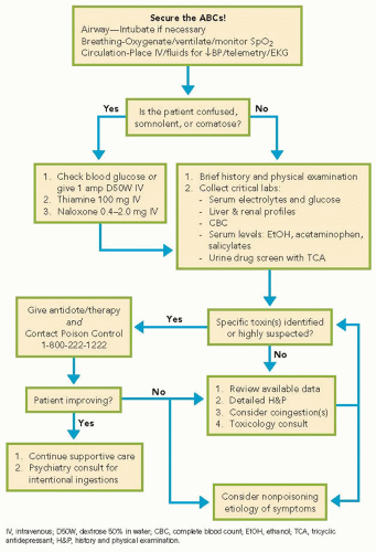


TABLE 33.4 Specific Antidotes for Selected Toxins


Toxin


Antidotes


Acetaminophen


N-acetylcysteine


Carbon monoxide


100% O2, hyperbaric O2 in some cases


Cholinesterase inhibitors (e.g., organophosphates)


Atropine 1-5 mg IV, repeat q5-10 min for ongoing wheezing or bronchorrhea


Pralidoxime 1-2 g IV over 30 min, repeat after 1 hr if ongoing weakness or fasciculations


Cyanide


Sodium nitrite 300 mg IV over 2-5 min


Digoxin


Digoxin-specific antibody fragments (Fab)


Acute: 10-20 vials Chronic: 3-6 vials


Ethylene glycol


Fomepizole 15 mg/kg IV over 30 min (first dose), then 10 mg/kg q12h × 4 doses, then 15 mg/kg q12h as needed


Iron Deferoxamine start at 5 mg/kg/hr, titrate as tolerated to 15 mg/kg/hr, max daily dose 6-8 g/day


Isoniazid


Pyridoxine 1 g for each gram of isoniazid ingested up to 70 mg/kg or 5 g max infused IV at 0.5 g/min until seizures stop, then the remainder infused during 4-6 hr


Methanol


Fomepizole 15 mg/kg IV over 30 min (first dose), then 10 mg/kg q12h × 4 doses, then 15 mg/kg q12h as needed


Methemoglobinemia


Methylene Blue 1-2 mg/kg IV over 5 min followed by 30 mL saline flush


Opioids


Naloxone 0.4-2 mg IV (IM, SC, or endotracheally)


Sulfonylureas


Octreotide 50 mcg SC q6h


IV, intravenously; IM, intramuscularly; SC, subcutaneously.


TREATMENT STRATEGIES


Antidotes

Specific antidotes are available for relatively fewtoxins. Although potentially life-saving, many of these antidotes have adverse effects and can be harmful if used inappropriately. Consultation with a poison control center or a medical toxicologist is advised when prescribing an antidote with which one is unfamiliar. A select list of antidotes is included in Table 33.2.


Gastric Decontamination

Among methods for blocking the absorption of drugs in the gastrointestinal (GI) tract, activated charcoal and whole-bowel irrigation (WBI) are recommended for use in limited circumstances. The routine use of gastric lavage, induced emesis (e.g., with syrup of ipecac), and cathartics are not recommended.

Activated charcoal, given orally or through a nasogastric tube, readily adsorbs most toxins, thereby preventing systemic absorption and toxicity. Substances that are not well adsorbed by activated charcoal are alcohols, iron, and lithium. The efficacy of activated charcoal is greatest when given within 1 hour of ingestion. A single dose of activated charcoal (1 g/kg) is recommended in adolescents and adults. Multiple-dose activated charcoal (MDAC) may be used as means of drug elimination in the management of certain poisonings (see below). Contraindications to the use of activated charcoal include an unprotected airway and the ingestion of a hydrocarbon. Caution should be taken in the setting of significant GI pathology or recent GI surgery.

WBI involves the enteral administration of large volumes of an osmotically balanced polyethylene glycol electrolyte solution to induce diarrhea with rapid expulsion of unabsorbed toxins from the GI tract. No controlled studies have been published, but WBI may be considered in the management of certain ingestions (Table 33.5). WBI is best performed using a nasogastric tube, and a recommended regimen is 1500 to
2000 mL/hr of enterally administered WBI fluid continued at least until a clear rectal effluent is noted. Contraindications include an unprotected airway, bowel perforation or obstruction, ileus, significant GI hemorrhage, toxic colitis, uncontrolled vomiting, and hemodynamic instability.


TABLE 33.5 Potential Indications for Whole-Bowel Irrigation


Iron poisoning


Lithium poisoning


Sustained-release or enteric coated medication toxicity


Retained illicit drug packets (i.e., from “body packing”)


Contraindications


Bowel obstruction


Ileus


Bowel perforation


Unprotected airway


Uncontrolled vomiting


Hemodynamic instability


Toxic colitis


Only gold members can continue reading. Log In or Register to continue

Stay updated, free articles. Join our Telegram channel

Oct 20, 2016 | Posted by in CRITICAL CARE | Comments Off on Toxicology

Full access? Get Clinical Tree

Get Clinical Tree app for offline access