Three- and Four-Dimensional Ultrasound in Neuraxial Anesthesia
David Belavy
There is scant literature on the use of three-dimensional (3D)/four-dimensional (4D) ultrasound in regional anesthesia and only one publication that explores the potential use of 4D ultrasound for neuraxial procedures.1, 2, 3, 4, 5, 6, 7, 8, 9, 10, 11, 12and 13
Neuraxial 4D ultrasound builds on the knowledge of 2D ultrasound scanning procedures, sonoanatomy, and needle technique. There are potential uses in preprocedural scanning and for needle guidance. The use of 4D ultrasound greatly depends on how the images are presented to the operator.
This chapter discusses how 3D/4D ultrasound data is acquired and presented, how the spine appears using this technology, and how it could be applied to epidural and spinal techniques.
3D/4D image acquisition
In two-dimensional (2D) ultrasound imaging, a 3D appreciation of the anatomy is built in the sonographer’s mind by examining structures in multiple 2D views, or sections, acquired through movement of the probe. Abdominal and obstetric curvilinear low-frequency transducers (2 to 5 MHz) are recommended for neuraxial imaging in adults.14 Their low frequency improves imaging of deeper structures, and curved probes provide a wider field of view. These 2D ultrasound probes typically consist of a single line of up to 256 piezoelectric crystals, which examine a 2D area and produce a B-mode image. The two dimensions are the depth and width of the imaging field. In contrast, static 3D ultrasound captures a volume of data that may then be digitally manipulated and displayed without movement of the probe. 3D imaging adds an elevation dimension to the depth and width of 2D imaging to describe a 3D volume of data. Each data element, or voxel, represents the echogenicity of that point in the 3D volume of tissue.
For the purposes of neuraxial ultrasound, there are two methods that 3D/4D ultrasound systems use to examine a 3D volume.15
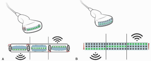
Mechanically steered arrays. In these systems, a sTandard curvilinear transducer with a single line of piezoelectric crystals is contained within a fluid-filled housing. The transducer is mechanically steered within the housing and sweeps over the area examined, building a volume of data (Fig. 7.1A). The time taken to acquire one volume of data is the time taken for the transducer to cover the sweep angle.

Figure 7.1. A: Image acquisition from a mechanically steered transducer. B: Image acquisition from an electronically steered transducer.
2D phased array transducer. In contrast to conventional transducers that contain a line of piezoelectric crystals, these transducers contain a 2D array of crystals. From this array, the ultrasound beam is electronically swept through the volume being examined, resulting in a more rapid examination. These transducers require more piezoelectric crystals; an example is the Philips X6-1 xMATRIX Array transducer which contains 9,212 elements (Fig. 7.1B).
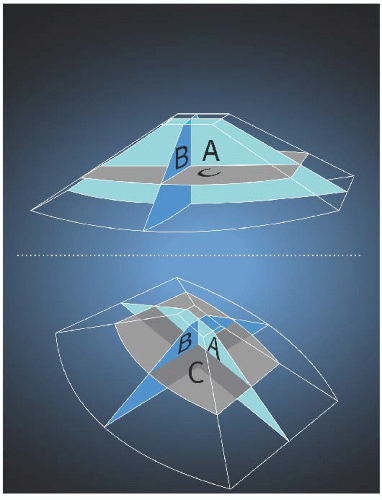
Both techniques produce data covering a pyramidal volume (Fig. 7.2). Real-time 3D (RT3D) or 4D ultrasound simply refers to repeated acquisition of 3D ultrasound volumes over time, producing a real-time examination. The image resolution and the dimensions of the volume of interest (imaging depth, sector width, and sweep angle) are key determinants of the frame rate in 4D ultrasound imaging.
3D/4D image display
The data acquired during a 3D scan can be examined on the ultrasound machine or off-line on a workstation. The two main postprocessing techniques relevant for displaying neuraxial images from a 3D dataset are multiplanar reconstruction (MPR) and 3D rendering.
Multiplanar reconstruction
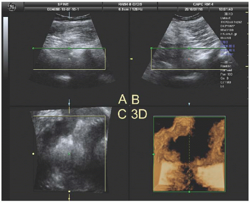
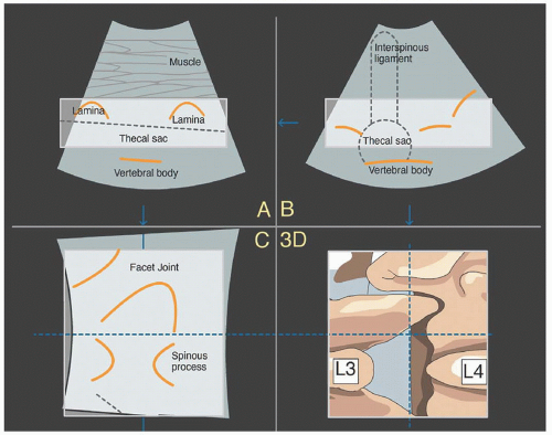
MPR allows any slice taken from a 3D dataset to be displayed, but initially, three orthogonal planes are chosen. MPR images are routinely displayed from spiral computed tomography (CT) and magnetic resonance imaging (MRI) scans in coronal and sagittal planes in addition to the traditional transverse or axial plane. Similarly, with 3D ultrasound, two or three orthogonal images are displayed from the acquired data (Fig. 7.3). The images of the three planes are usually designated A, B, and C. The first plane is the familiar 2D ultrasound image. Plane B or y is perpendicular to the first. If plane A is positioned over the long axis of a structure, plane B would display its short axis. Plane C or z is perpendicular to the other images and represents echoes from a plane positioned a disTance from the probe. Remembering the terms can be aided by using the mnemonic “Actual, Bisecting, and Coronal.” Figures 7.3 and 7.4 demonstrate these views at the lumbar vertebral level.
 Figure 7.3. A 3D sonogram of the right paramedian L3/L4 vertebral interspace. A: Longitudinal paramedian image. B: Axial image. C: Coronal image. 3D: 3D reconstruction. |
 Get Clinical Tree app for offline access 
|


