(1)
Thoracic and Vascular Trauma, R Adams Cowley Shock Trauma Center, Baltimore, MD, USA
Keywords
Thoracic vascular traumaPenetrating injuryBlunt injuryBlunt aortic injury (BAI)Thoracic endovascular aortic repair (TEVAR)Delayed operative managementSelective nonoperative managementComputed tomographic angiography (CTA)ShockHemodynamic instabilityClamshell thoracotomyMedian sternotomyIntroduction
Thoracic vascular trauma can present a significant clinical challenge. The relative inaccessibility of the vasculature, exsanguinating hemorrhage, and lack operator experience all contribute to the difficulty in treating these infrequent but potentially lethal injuries. Early recognition, appropriate utilization of imaging, and familiarity with surgical exposure and techniques of repair will improve survival. This chapter includes a brief history of thoracic vascular trauma, the evolution of treatment, specific operative techniques, management of surgical complications, and outcomes. Emphasis will be placed on decision making, clinical judgment, and operative techniques.
History
Although thoracic vascular trauma is not frequent, it is associated with significant mortality. The vast majority of thoracic vascular injuries are the result of penetrating trauma, all intrathoracic vessels are at risk, and the mortality is significant [1–5]. Blunt injuries most often involve the descending thoracic aorta just distal to the left subclavian artery and are also associated with high mortality [6]. Several studies report a prehospital mortality greater than 50 % following penetrating injury [1, 7] and a variable operative mortality ranging from zero to almost 40 % [8]. Parmley’s landmark article described the natural history and lethality of blunt aortic injury (BAI) [6]. Unlike penetrating trauma, there have been major advances in the treatment and survival of BAI, including thoracic endovascular aortic repair (TEVAR), delayed operative management, and selective nonoperative management. Endovascular techniques have been less frequently employed in the treatment of penetrating vascular injuries. Improved imaging, particularly computed tomographic angiography (CTA), defines the nature, extent, and location of the vascular injury. This information influences the decision regarding nonoperative, endovascular, or open procedure. By characterizing the injury and its location, imaging will influence the choice of incision. Damage control, a widely used technique for selective abdominal and orthopedic injuries, has been successfully applied to both vascular and thoracic trauma.
Penetrating Vascular Injury
The thorax, especially the mediastinum, contains several large arteries and veins. The aorta and its intrathoracic branches and the innominate, subclavian, and proximal carotids can all be injured from blunt or penetrating injury. Intrathoracic venous structures including the superior and inferior vena cavae and right and left innominate, subclavian, proximal internal jugular, and azygos veins are all at risk. The pulmonary artery and veins may also be injured, especially at the pulmonary hilum.
The vast majority of the thoracic great vessel injuries result from penetrating trauma, and many victims die before reaching definitive care [9]. Patients arriving with suspected great vessel injury demand rapid assessment and evaluation. Not surprising, those presenting in shock generally have a higher mortality, reinforcing its lethality. This subset of patients requires an immediate operation; only stable patients should undergo advanced imaging.
Initial Evaluation
All patients presenting with penetrating thoracic trauma are at risk for great vessel injury and undergo the standard ABCs of trauma care. Vital signs, arterial saturation, and a rapid yet thorough physical exam, with particular attention to external bleeding, expanding hematomas, and upper extremity pulse differential, should be performed. Direct digital pressure should be applied to external bleeding. An absent radial pulse suggests an arterial injury, and an extremity neurologic deficit may be the result of a brachial plexus injury. A FAST examination including abdominal and pericardial views will detect hemoperitoneum and hemopericardium. A portable chest radiograph yields valuable information including hemothorax, pneumothorax, or widened mediastinum. A type and cross match and lactate and arterial blood gas should be included with the admission laboratory tests. With these data the clinician must make a judgment regarding hemodynamic stability, shock, and depth of shock. Hypotension, generally defined as a systolic blood pressure <90 mmHg, is an ominous sign with resultant shock and acidosis. A normal blood pressure on admission may represent a compensated shock state, which should be considered in the presence of tachycardia and a narrow pulse pressure. Lactate and base deficit are excellent markers for the presence and depth of shock.
The initial determination of shock and hemodynamic instability is the crucial first decision in the patient’s management. Unstable patients require immediate operative intervention. Additional diagnostic studies and imaging should only be performed in stable patients. This point cannot be overemphasized; penetrating thoracic trauma with hemodynamic instability and shock requires surgery.
Hemodynamically stable patients, however, may benefit from additional imaging. CTA has supplanted angiography as the imaging modality of choice for both blunt and penetrating chest trauma. It defines the nature, extent, and exact location of the injury. Information gleaned influences open versus endovascular approach and, choice of incision if an open repair is indicated. CTA accurately diagnoses penetrating great vessel injuries, altering the operative approach in 25 % of patients [8]. Chest CTA has been shown to be the definite imaging study for penetrating mediastinal injuries, which historically required angiography, bronchoscopy, esophagoscopy, and esophagram to exclude an injury [10]. Blunt aortic injury is also accurately diagnosed with CTA [11].
Surgical Approach
The choice of the incision (“the incision decision”) requires sound surgical judgment and is influenced by the patient’s hemodynamics. Unlike abdominal exploration, which is almost always performed through a laparotomy incision, there are several surgical approaches to the thorax, each with advantages and disadvantages. Irrespective of the choice of incision, it must provide adequate surgical exposure and be versatile. The thorax can be explored through an anterolateral incision, which can be extended across the midline as a bilateral thoracotomy or “clamshell” (Figs. 18.1 and 18.2). This offers several advantages; it is rapid, allows excellent exposure of the anterior mediastinum and pleural spaces, and is an incision familiar to trauma surgeons. With the patient supine, a laparotomy can be easily performed. The disadvantages include inadequate access to posterior structures, and, if a clamshell is performed, the incision across the sternum may be placed too caudal, limiting superior mediastinal exposure. Placing a bump under the back and extending the ipsilateral arm improve exposure. When performing a clamshell thoracotomy, remember to ligate the internal mammary arteries. With profound hypotension they may not initially be bleeding but certainly will once blood pressure is restored.
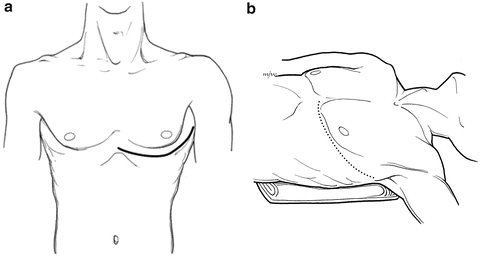
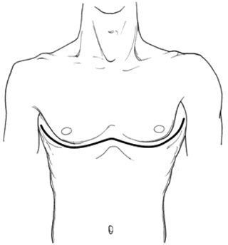

Fig. 18.1
(a and b) Anterolateral thoracotomy. The bump and extended ipsilateral arm greatly improves exposure of the pleural space. The incision can be extended across the sternum as a bilateral anterolateral thoracotomy (“clamshell”).

Fig. 18.2
Anterolateral thoracotomy extended as a clamshell. For optimal exposure the sternum must be divided as shown. Placing the incision more inferiorly will transect the xiphoid thereby limiting exposure to the superior mediastinum.
Median sternotomy is an excellent choice for mediastinal exposure (Fig. 18.3). It is ideal for cardiac and great vessel injury, can be extended for neck or periclavicular exposure, and allows a laparotomy to be easily performed. A surgeon familiar with this incision can rapidly perform it, but less experienced operators may prefer the clamshell approach. As with the anterior lateral incision, sternotomy provides poor visualization of posterior structures. One area of controversy is the approach to the proximal left subclavian artery. While the right subclavian is exposed through a sternotomy with periclavicular extension, the optimal exposure of the left subclavian artery has been debated. Conventional wisdom favors a periclavicular incision coupled with a third or fourth interspace left anterior thoracotomy. This is curious since the operative approach to the proximal left carotid artery is median sternotomy and the left subclavian vessel is adjacent (Fig. 18.4). Combining sternotomy with a left supraclavicular extension provides excellent exposure of the left subclavian artery [12]. We have used this approach exclusively for left subclavian injuries, and clavicular resection is rarely needed [8]. Hemodynamically unstable patients require rapid evaluation, a thoughtful choice of incision and prompt surgical exploration. Anterolateral thoracotomy, clamshell, and sternotomy are all acceptable incisions in the hemodynamically unstable patients [8].
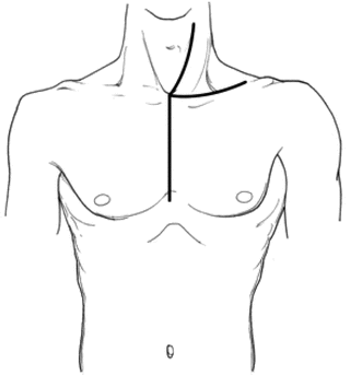
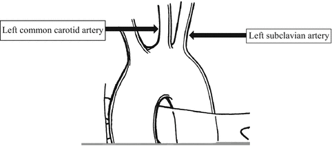

Fig. 18.3
Median sternotomy. It is the ideal approach to the heart and great vessels. This is a versatile incision as it can be extended to the neck or clavicle.

Fig. 18.4
Aortic arch and branches. The left subclavian artery is adjacent to the left common carotid and can be exposed through a sternotomy with left periclavicular extension. This is the authors’ preferred approach.
There are more options for surgical exposure in the hemodynamically stable patient with a great vessel injury. As opposed to an emergent operation, which is commonly exploratory in nature, imaging studies will have defined the location and nature of the vascular injury. The operation is performed to repair a definitive injury, thus allowing a more tailored approach. In addition to anterolateral thoracotomy (unilateral or clamshell) and sternotomy, periclavicular, partial sternotomy and posterolateral thoracotomy approaches can be used. The choice of incision depends on the specific injury, and each has its inherent advantages and disadvantages.
A periclavicular incision has the advantage of being rapidly performed and is versatile, as it can be extended to a sternotomy or for a neck exploration (Fig. 18.5). The main disadvantage is limited exposure, and clavicular resection is necessary and may prove more challenging than anticipated, especially for those with limited experience with the technique. Posterolateral thoracotomy allows excellent visualization of the pleural space with the exception of the anterior mediastinum and is the preferred incision for elective thoracic surgery (Fig. 18.6). Disadvantages, in addition to the limited anterior exposure, include lack of versatility. Also hypotension may be exacerbated with lateral positioning. Single-lung ventilation will allow for excellent visualization of the pleural space. In stable patients partial sternotomy is an attractive option for superior mediastinal exposure (Fig. 18.7). The manubrium is divided in the midline from the sternal notch passed the angle of Louis. It is carried laterally as a “T” of “J” and a small sternal retractor (pediatric sternal retractors work well) is placed. It affords excellent exposure of superior mediastinum and is versatile, as it can be extended to the neck and clavicle or continued as a full sternotomy. The ultimate choice of incision for exploring the hemodynamically stable patient will depend on the location of the injury, experience, and surgical judgment.
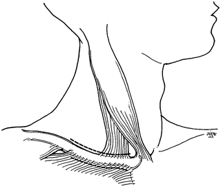
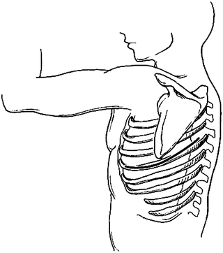
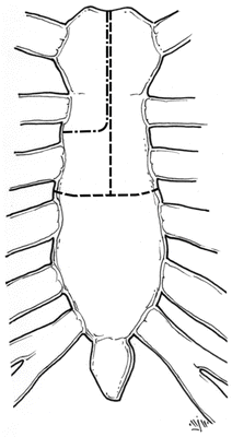

Fig. 18.5
Periclavicular incision. Either supra- or infraclavicular approach can be used. Resecting the clavicle may be challenging and take more time than expected. Unroofing a hematoma with proximal control can be problematic.

Fig. 18.6
A posterolateral approach is preferred for elective thoracic operations. Limited exposure of the anterior mediastinum and lack of versatility are its disadvantages. Exposure of the hemithorax is greatly improved with double lumen tube and lung isolation.

Fig. 18.7
Partial sternotomy. This is an attractive option in stable patients. Carrying the sternotomy distal to the angle of Louis generally provides excellent exposure to the superior mediastinum. Either a “T” or “J” lateral division of the sternum can be utilized. This incision is versatile as it can be extended to the neck and clavicle or continued as a full median sternotomy.
Intraoperative Management
Operative management of intrathoracic vascular trauma depends on the specific vessel injured and the patient’s clinical condition. Damage control is an attractive option in the hypotensive, acidotic, hypothermic, and coagulopathic patient. Hemorrhage is controlled, the thorax is packed, and resuscitation continues in the ICU. Following restoration of normal physiology, planned re-exploration and closure can be performed [13]. Proximal and distal vessel control is a fundamental tenant of vascular surgery; the previous section discussed the various operative approaches to accomplish this. With the expanding role of catheter-based therapy, a sophisticated option is proximal balloon occlusion. This is an excellent, though underutilized, technique to achieve inflow control without extensive dissection in a challenging anatomic location. Its role will expand as catheter-based therapies gain more popularity.
Although thoracic venous injuries may be repaired, it can be time consuming, often results in venous thrombosis and possible embolization. With the crucial exception of the superior and inferior vena cavae, which are repaired without lumen compromise, thoracic veins can be ligated with little clinical consequence. If ligation is performed, indwelling venous catheters must be removed prior to venous ligation. Facial or extremity edema is the sequela of venous ligation, which is managed by elevation and generally resolves within days.
With few exceptions arterial injuries should be repaired, with ligation only performed as life-saving measure for uncontrollable ensanguining hemorrhage. There are multiple options for arterial repair, and the trauma surgeon should be familiar with all of them. They include primary repair, patch angioplasty, graft interposition, and temporary shunting with delayed repair. Stab wounds can often be repaired primarily, with or without resection as indicated. Patch angioplasty can be accomplished with autologous vein or prosthetic material. Gunshot wounds result in significant tissue injury and generally necessitate resection and end-to-end interposition graft. Contrary to the use of bypass grafting for diffuse atherosclerotic disease, penetrating injuries are localized and amenable to graft interposition. Either autologous vein or prosthetic graft can be used. Size match and wound contamination will influence the choice of conduit. Vein is the preferred conduit in a grossly contaminated wound or if the graft traverses a joint.
Stay updated, free articles. Join our Telegram channel

Full access? Get Clinical Tree


