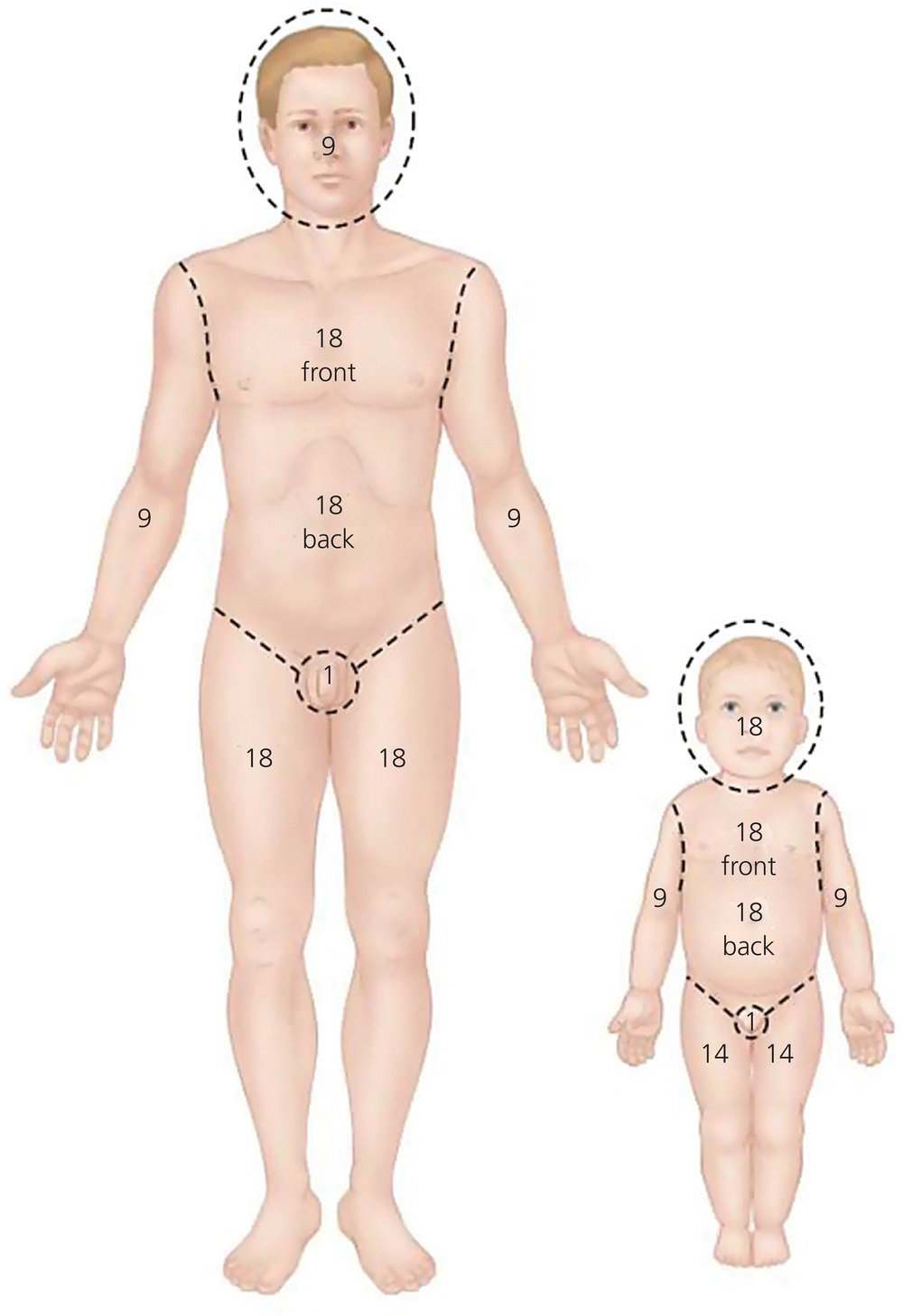
Chapter 33 John McManus, Richard B. Schwartz, and Sabina A. Braithwaite A contemporary understanding of burn injuries is essential for all out-of-hospital providers. Burn injuries carry high morbidity and mortality, resulting in severe pain, scarring, and permanent disability. Additionally, specialized resources such as burn centers are required for care and recovery. Deaths from fires and burns are the third leading cause of fatal home injury. The US burn mortality rate ranks eighth among the 25 developed countries [1]. According to the American Burn Association National Burn Repository 2012 statistics, over 450,000 victims received medical treatment for burns in the US in the last decade [1]. The majority of these burns result from fire and/or flame injuries and contact with hot objects. Chemical burns account for approximately 3% of burns and 7% of burn admissions annually. Approximately 3,400 deaths occurred (most from smoke inhalation), including 2,550 deaths from residential fires (most from cooking), 300 from vehicle crash fires, and 550 from other sources (approximately 150 deaths from flame burns or smoke inhalation in non-residential fires, 400 from contact with electricity, scalding liquids, or hot objects). Although the number of fatalities and injuries from residential fires has declined gradually, many residential fire-related deaths remain preventable and pose a significant public health problem. Over 60% of US acute burn hospitalizations were admitted to 127 burn centers [1]. Such centers each average over 200 annual admissions for burn injury and skin disorders requiring similar treatment. The other 4,500 US acute care hospitals average fewer than three burn admissions each per year [1–4]. Fire and burn injuries represent 1% of the incidence of injuries and 2% of the total costs of injuries, or $7.5 billion each year [5]. Risk factors for burn injuries include extreme age groups (<4 years and >65 years), poverty, African and Native American descent, and rural area dwellers. Most adults have sustained burns during their lives. The skin is the largest organ in the body and serves as a barrier to outside insults and injuries. The skin protects against water loss, entrance of undesirable substances (microorganisms, toxins), mechanical shock and forces, extreme environmental temperatures, and ultraviolet light damage to keratin and melanin. Furthermore, the skin is involved in sensory perception, temperature regulation, and biochemical activities (e.g. vitamin D synthesis). The skin is made up of three basic layers. The outer layer, the epidermis, is the thin outer layer of the skin which consists of the stratum corneum containing fully mature keratinocytes which produce fibrous proteins (keratins) that are continuously shed (prevents the entry of most foreign substances as well as the loss of fluid from the body), the keratinocyte layer containing living keratinocytes (squamous cells), and the basal layer, the deepest layer of the epidermis, containing basal cells (continually dividing and forming new keratinocytes). The middle layer of the skin, the dermis, contains blood vessels, lymph vessels, hair follicles, sweat glands, fibroblasts, and nerves. The dermis is held together by collagen, made by fibroblasts, and gives skin flexibility and strength. The dermis also contains pain and touch receptors. The subcutis is the deepest layer of skin and consists of a network of collagen and fat cells that aid in conserving the body’s heat and protect the body from injury by acting as a “shock absorber.” Accurate assessment of the burn patient and appropriate institution of early care are critical to optimal outcomes. Although burn size and depth are obvious factors in determining burn severity, the location (body part) of the burn, age of the patient, preexisting disease, and presence of trauma, including inhalation injury, may complicate treatment. Specific anatomical locations of burns often result in significant morbidity and mortality disproportionate to burn size (i.e. head, neck, hands, feet, perineum, and genitalia). Furthermore, patients <2 years or >50 years old are at higher risk of complications and death than the remaining population [1]. In infants, thin skin, limited reserves, and high surface area-to-mass ratios contribute to this risk, whereas thinning skin and medical problems commonly associated with aging are major factors in older individuals. Young children are also at risk for burns caused by abuse. These injuries are most often scald burns from tap water, are deeper than those seen in the general pediatric burn population, and commonly involve the lower extremities, buttocks, and genitalia. Pediatric and elderly burns may often be an initial presentation of abuse and should be considered in the differential diagnosis. There are several ways to classify burns (depth, severity, and surface area). Burn depth is a product of temperature, duration of exposure, and skin thickness, with depth being described in its relationship to total skin thickness. Most burns have areas that are of mixed depth, with deeper burns often occurring in areas of thinner skin. The older classification of describing “degrees” of burn is not often used any more. Rather, the American Burn Association now uses the total body surface area and the severity (partial verses full thickness) of injury as a modern descriptor (Tables 33.1, 33.2). The old descriptive terms are paired with the newer classification system in order to understand the changes. Table 33.1 Classification of burns based on depth Table 33.2 American Burn Association classification of burns by total body surface area (TBSA) First-degree (superficial) burn injuries involve only the epidermis or topmost layer of skin and are recognized by their erythematous appearance and lack of blisters or skin separation. The classic first-degree injury is the sunburn or superficial scald burn from spills. These burns usually have morbidity restricted only to pain, and are therefore not classified into burn size. Second-degree (superficial or deep partial thickness) burn injuries involve the epidermis and part way through the dermis. Epithelial elements remain in the undestroyed dermal appendages and spontaneous healing usually occurs in 7–28 days. Second-degree burns are very painful and are usually blistered. Third-degree (full-thickness) burn injuries are those that extend through the dermis, destroying all epidermal and dermal elements. They may initially have blisters containing hemorrhagic fluid and/or dead tissue (eschar). The presence or absence of pain is an unreliable indicator of depth and severity. Accurate initial assessment of burn size is essential for optimal patient care. Burn size is expressed as total body surface area (TBSA) or body surface area (BSA), where approximately 1% of a patient’s surface area is equal to the palmar surface of the patient’s hand with the fingers closed. This measurement is most useful for small (<5% TBSA) or spotty burns. For larger areas, the rule of nines (Figure 33.1) for adults provides a simple and rapid estimation of burn size in the adult. Figure 33.1 Rule of nines. Source: Dorland’s Illustrated Medical Dictionary, 32nd edn. Philadelphia: Elsevier Saunders, 2011. Reproduced with permission of Elsevier. When calculating burn size using any method, first-degree burns are not counted and only the proportion of area with at least a partial-thickness burn is calculated. Thus, for an upper extremity to be considered 9% TBSA, the entire extremity from the shoulder to the finger tips must be burned at least to the blistering level. If only the posterior half of the upper extremity is burned, then burn size is considered to be 4.5% TBSA. Calculating pediatric burns is often challenging and can be inaccurate if the provider is not appropriately trained. The rule of nines (see Figure 33.1) has also been used for pediatric patients. However, the Lund and Browder classification can also be used to more precisely calculate the percentage of BSA burned by mapping the injured areas of the body on charts detailing age-appropriate measurements. This method identifies the different body proportions according to the age of the patient (with children having larger heads and smaller lower extremities than adults) and through dividing the body into smaller units, such as dividing the upper extremity into the upper arm, lower arm, and hand. Computer programs are now being used to estimate surface area calculations. Inhalation injury is a complex set of pathophysiological reactions that occur from exposure to smoke and/or chemical products. Systemic and respiratory damage can result in significant morbidity and mortality as well as permanent dysfunction [6,7]. When combined with thermal injury, inhalation injury increases pulmonary compliance and fluid requirements, and doubles mortality. Technically, injury is a misnomer, and inhalation injury is really the result of fluid shifts caused by external burns. These conditions do not necessarily imply pulmonary injury, because they also occur with scald and chemical burns. Edema formation in the posterior pharynx and glottic and subglottic areas associated with deep burns of the upper chest, neck, and lower face has the potential to occlude the upper airway. Tachypnea and stridor are often late signs and when absent are unreliable in ruling out airway injury. Airway injury is diagnosed by fiberoptic bronchoscopy [8]. Early grading of inhalation injury severity is often inaccurate. The injury is basically a chemical burn from which resulting edema of the small airways creates distal microatelectasis and a clinical picture identical to acute respiratory distress syndrome. Lower airway or “smoke inhalation” injury is caused by the patient inhaling the products of combustion, often as a result of being in a confined space. Specific injuries resulting from specific toxins, cyanide and carbon monoxide, are discussed elsewhere in this text.
Thermal and chemical burns
Introduction
Pathophysiology
Severity
Depth
Classification
Cause
Appearance
Sensation
Healing time
Scarring
Superficial burn
Ultraviolet light, very short flash (flame exposure)
Dry and red; blanches with pressure
Painful
3–6 days
None
Superficial partial-thickness burn
Scald (spill or splash), short flash
Blisters; moist, red and weeping; blanches with pressure
Painful to air and temperature
7–20 days
Unusual; potential pigmentary changes
Deep partial-thickness burn
Scald (spill), flame, oil, grease
Blisters (easily unroofed); wet or waxy dry; variable color (patchy to cheesy white to red); does not blanch with pressure
Perceptive of pressure only
More than 21 days
Severe (hypertrophic) risk of contracture
Full-thickness burn
Scald (immersion), flame, steam, oil, grease, chemical, high-voltage electricity
Waxy white to leathery gray to charred and black; dry and inelastic; does not blanch with pressure
Deep pressure only
Never (if the burn affects more than 2% of the total surface area of the body)
Very severe risk of contracture
Type of burn
Minor
Moderate
Major
Criteria
<10% TBSA burn in adult
10–20% TBSA burn in adult
>20% TBSA burn in adult
<5% TBSA burn in young or old
5–10% TBSA burn in young or old
> 10% TBSA burn in young or old
<2% full-thickness burn
2–5% full-thickness burn
> 5% full-thickness burn
High-voltage injury
High-voltage burn
Suspected inhalation injury
Known inhalation injury Any significant
Circumferential burn
burn to face, eyes, ears, genitalia or joints
Concomitant medical problem predisposing the patient to infection (e.g. diabetes, sickle cell disease)
Significant associated injuries (e.g. fracture, other major trauma)
Disposition
Outpatient management
Hospital admission
Referral to burn center
Burn size
Inhalation injury
Related posts:


Stay updated, free articles. Join our Telegram channel

Full access? Get Clinical Tree


