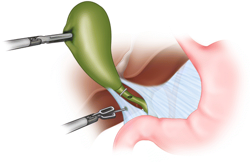
Fig. 5.1
A catheter is introduced into the abdomen through a separate subcostal stab incision via an angiocatheter or sheath. A clip is placed at the gallbladder/cystic duct junction and a ductotomy made in the cystic duct through which the cholangiocatheter is advanced
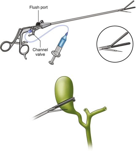
There are several commercially available devices that can be utilized to facilitate cholangiography. The Olsen Cholangiogram Clamp (Karl Storz Endoscopy, Culver City, California) is an instrument that can be used to grasp the cystic duct and allows passage of a 4- to 5-French ureteral catheter into the cystic ductotomy (Fig. 5.2). This instrument can be passed through either the subcostal or the epigastric port and therefore does not require an additional incision for introduction of a cholangiocatheter. As an alternative to IOC via a cystic ductotomy, imaging of the biliary system can be performed via a catheter inserted into the gallbladder. The Kumar Pre-View Clamp (Nashville Surgical Instruments, Springfield, Tennessee) is a locking grasper placed across the gallbladder fundus with a channel through which a catheter with a 1.25 cm 19 gauge needle can be passed and advanced into the Hartmann’s pouch of the gallbladder for contrast injection (Fig. 5.3) [16]. The clamp can be introduced into the abdomen via the subcostal port. This method has a theoretical advantage of not requiring an incision in any ductal structures. Thus, this may be preferable in patients with a short cystic duct or where the ductal anatomy is unclear. However, this method may not be feasible when there is a stone obstructing the neck of the gallbladder or the cystic duct unless the stone can be dislodged.


Fig. 5.2
The Olsen clamp can be placed through either the mid-subcostal or the epigastric port. A clip is still placed at the gallbladder/cystic duct junction, but no additional clip is necessary to hold the catheter in place. The catheter is threaded through a channel in the clamp and is held in place via the clamp on the cystic duct

Fig. 5.3
(a) Kumar Pre-View Clamp has a channel through which the cholangiocatheter with a needle can be advanced into the Hartmann’s pouch of the gallbladder. Contrast can be injected via the cholangiocatheter. (b) The clamp can be applied across the fundus to prevent contrast from flowing retrograde into the gallbladder
There has only been one randomized trial comparing the Olsen to the Kumar clamp for IOC. There were no differences between the two clamps in terms of success rate in obtaining the IOC, the mean IOC time, or surgeon perception of the ease of using the clamps [17]. However, the trial only included 59 laparoscopic cholecystectomy cases and surgeons had greater familiarity with the Olsen clamp. Given the relative advantages and disadvantages of each method, it is important for surgeons to learn and become proficient with each method in order to be able to perform IOCs across different presentations of acute cholecystitis or other biliary disease.
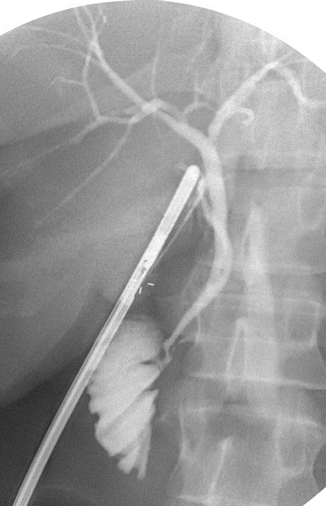
Once the cystic duct or gallbladder fundus has been cannulated, then fluoroscopy can be performed. During the initial infusion of contrast, 3–5 mL of dye is injected and the cystic duct/bile duct junction is observed. Attention should be paid to the length of the cystic duct and whether or not it contains any stones. The angle of insertion of the cystic duct into the common or right hepatic duct and any anatomic abnormalities should be noted as they may increase likelihood of bile duct injury during cholecystectomy. As the dye is injected, the entire biliary tree should be visualized to the third level of the intrahepatic ducts. A normal cholangiogram should demonstrate standard intrahepatic and extrahepatic ductal anatomy without anatomical variations or ductal dilation; there should be normal tapering of the bile duct towards the sphincter of Oddi with prompt passage of contrast into the duodenum (Fig. 5.4). The biliary tree should be examined for any aberrant ductal anatomy that may cause a predisposition to a bile duct injury such as a short cystic duct, cystic duct insertion into the right or left hepatic ducts, an accessory right hepatic duct, or an accessory cystic duct.


Fig. 5.4
A normal intraoperative cholangiogram showing filing of the intrahepatic and extrahepatic ducts, narrow tapering of the common bile duct, no filling defects, and emptying of contrast into the duodenum
Biliary ductal dilatation should prompt investigation for a cause of bile duct obstruction such as choledocholithiasis demonstrated by intraluminal filling defects, an extraluminal stricture, or non-filling of the duodenum. If the duodenum does not easily fill with contrast, there may be a distal obstructing common bile duct stone, biliary stricture, or spasm of the sphincter of Oddi. If sphincter spasm is suspected, 1 mg of glucagon can be infused intravenously to cause relaxation of the sphincter and the cholangiogram can then be repeated. If dye does not freely pass with this maneuver, a common bile duct stone or biliary stricture should be suspected.
Evidence for Routine Versus Selective IOC
IOC and CBDI
The primary reason for promoting routine IOC during laparoscopic cholecystectomy is the prevention of CBDI. Multiple cohort and case–control studies have examined the use of IOC. In 2002, 40 case series comprised of 327,523 laparoscopic cholecystectomies were analyzed [18]. The authors determined that there was an association between routine IOC use and a lower incidence of CBDI (0.21 % versus 0.43 %) and a higher rate of diagnosis at the time of initial operation (87 % versus 44.5 %). However, these data may be biased due to flaws in the study designs such as lack of standardized definitions for CBDI and selection bias in the performance of IOC.
Several randomized controlled trials of IOC have been performed. A systematic review of the literature performed in 2012 identified eight randomized trials comprised of 1715 patients [3]. The incidence of CBDI was 0.2 % including cystic duct avulsions, and the incidence of major CBDI was 0.1 %. A meta-analysis to combine the data from the trials was considered inappropriate because of the low number of injuries, the poor quality of the trials, and considerable heterogeneity between trials. The authors concluded that the evidence from randomized trials neither supported nor refuted the effectiveness of routine IOC to prevent CBDI.
Given the increasing availability of large administrative databases, advanced statistical methods have been used to better define the association between IOC and CBDI [2, 4, 12, 15]. In 2001, Flum et al. published one of the first studies using statewide data [2]. Using 1991–1998 Washington hospital discharge data, they identified 76 major CBDIs out of 30,630 laparoscopic cholecystectomies for an overall incidence of 2.5 per 1000 operations; the incidence of CBDI decreased over the time period from 3.2 to 1.7 per 1000 operations. The authors identified a statistically significant 1.7-fold increased relative risk of CBDI when IOC was not used. Furthermore, they determined that less surgeon experience and decreased surgeon frequency of IOC use were significant predictors of CBDI.
A more recent analysis used statewide Medicare data. In 2013, Sheffield et al. examined 2000–2013 Texas Medicare data to define the association between IOC use and CBDI [15]. The authors identified 280 CBDIs out of 92,032 patients for an overall incidence of 0.3 %; 40.4 % of cases used IOC. Using traditional statistical analyses, nonuse of IOC was associated with a statistically significant, 1.8-fold increased odds of CBDI. Using advanced statistical methods to adjust for unmeasured confounders, they determined that there was no longer an association between IOC use and CBDI.
How should these data regarding the effectiveness of IOC for preventing CBDI be reconciled? The methodological limitations of case–control and cohort studies limit their utility, and the randomized trials were considerably underpowered to identify an effect of IOC on CBDI. The large database analyses provide significant advantages over these other study designs by providing more patients and therefore more power than all of the randomized trials combined. They also reflect “real-world” conditions rather than highly controlled circumstances as are present in randomized trials. Furthermore, use of advanced statistical methods can be used to infer whether or not a causal effect exists (i.e., whether use of IOC prevents CBDI) [19]. Nonetheless, limitations with these advanced methods must also be considered [20]. So, while the most recent evidence suggests that routine IOC is not an effective strategy for preventing CBDI, some caution should still be used in interpreting these results.
One unmeasured factor in large database analyses is the accuracy of individual surgeons in interpreting IOCs. Sanjay et al. evaluated IOC interpretation among 20 trainees and 20 fully trained surgeons [21]. They were asked to interpret 15 IOCs of normal anatomy as well as normal and abnormal variants of anatomy. Their accuracy was low for identifying normal anatomy and normal variants of anatomy, 45 % and 29.5 %, respectively. However, their accuracy for identifying abnormal anatomy was high at 95.5 %. There was no difference in accuracy based on trainee level or routine use of IOC. These findings are echoed by a case series of patients who had CBDIs; 43 % had IOCs and in two-thirds the injuries were not identified [22].
While the effectiveness of IOC to prevent CBDI has not been definitively proven, the cost-effectiveness of routine IOC to prevent CBDI has been debated. These cost-effectiveness analyses assume that IOC is an effective strategy. Even so, the answer regarding cost-effectiveness varies depending upon the estimated costs of IOC and the number of IOCs that need to be performed in order to prevent one CBDI. IOC is less cost-effective when the cost per IOC is higher and when the baseline risk of CBDI is lower (i.e., when performed by more experienced surgeons or during less complex cases) [2]. Using a cost per IOC of $122, Flum et al. estimated the cost of IOC per CBDI avoided was $87,100 (in year 2000 dollars) [2]. In contrast, using a cost per IOC of $700, Livingston et al. estimated the cost of IOC per CBDI avoided was $504,084 [23]. Using a cost of CBDI of $300,000 [23, 24], IOC is only cost-effective in preventing CBDI if the cost per IOC is low.
IOC and Suspected Choledocholithiasis
Another reason for performing IOC is to identify choledocholithiasis. While missed choledocholithiasis may result in recurrent episodes of biliary symptoms including complications such as gallstone pancreatitis or cholangitis, a systematic review of the literature suggested that the incidence of asymptomatic choledocholithiasis during laparoscopic cholecystectomy is only 4 % [25]. Furthermore, only 0.6 % of these common bile duct stones progress to symptoms. Another study where a catheter was left in place if common bile duct stones were found at IOC and imaging repeated in 6 weeks found that one-third of common bile duct stones passed spontaneously [26].
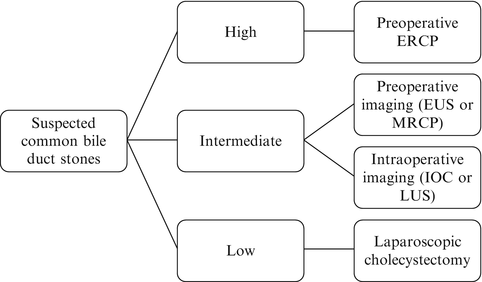
Multiple studies have been performed in order to identify predictors of choledocholithiasis given that preoperative risk stratification may alter the diagnostic and management algorithm in patients with symptomatic cholelithiasis or acute cholecystitis. The American Society for Gastrointestinal Endoscopy (ASGE) stratifies preoperative risk factors for choledocholithiasis into moderate, strong, and very strong and probability of choledocholithiasis into low, intermediate, and high risk (Fig. 5.5) [27]. For patients with a low probability of choledocholithiasis, they recommend laparoscopic cholecystectomy alone. For patients with a high probability of choledocholithiasis, they recommend ERCP. For patients with intermediate probability, there are multiple diagnostic and management strategies that are dependent upon local resources and expertise. These include preoperative endoscopic ultrasound (EUS) or magnetic resonance cholangiopancreatography (MRCP) followed by ERCP, laparoscopic cholecystectomy with IOC followed by common bile duct exploration or postoperative ERCP, and intraoperative laparoscopic ultrasound.


Fig. 5.5
Algorithm for managing suspected choledocholithiasis based on the presence of predictors. High suspicion is present if there is any strong predictor (common bile duct stone on transabdominal ultrasound, clinical ascending cholangitis, or bilirubin >4 mg/dL) or both strong predictors (dilated common bile duct on ultrasound defined as >6 mm and bilirubin 1.8–4 mg/dL). Intermediate suspicion is present if only one strong predictor or any moderate predictors are present (abnormal liver biochemical test other than bilirubin, age older than 55 years, and clinical gallstone pancreatitis). Low suspicion is present if no predictors are present. ERCP endoscopic retrograde cholangiopancreatography, EUS endoscopic ultrasound, MRCP magnetic resonance cholangiopancreatography, LUS laparoscopic ultrasound. From reference [27]
The cost-effectiveness of IOC to treat patients with suspected choledocholithiasis depends upon the probability of common bile duct stones. Urbach et al. performed a cost-effectiveness analysis comparing four strategies: preoperative ERCP followed by laparoscopic cholecystectomy, laparoscopic cholecystectomy with IOC and laparoscopic common bile duct exploration, laparoscopic cholecystectomy with IOC and postoperative ERCP, and laparoscopic cholecystectomy with expectant management [28]. Laparoscopic cholecystectomy with IOC and laparoscopic common bile duct exploration was the most cost-effective strategy, defined as the least cost per case of residual common bile duct stones prevented; this was true across probabilities of common bile duct stones. If the expertise is not available for laparoscopic common bile duct exploration, the authors recommended preoperative ERCP if the probability of common bile duct stones was greater than 80 %.
The cost-effectiveness of IOC in managing patients with suspected choledocholithiasis is derived in a large part from the high specificity of IOC. A prospective population-based study of over 1000 patients reported that IOC was feasible in 95 % of cases and had a sensitivity of 97 % and a specificity of 99 % for detecting choledocholithiasis [29]. With an incidence of 11 % of choledocholithiasis in that study, the negative predictive value was 99 % and the false negative rate was 1 %.
Brown et al. performed a decision and cost-effectiveness analysis in order to compare five strategies for treating suspected choledocholithiasis in patients with symptomatic cholelithiasis [30]. Using a specificity of 99 %, they determined that laparoscopic cholecystectomy with IOC followed by ERCP for positive findings was the most cost-effective strategy, defined as cost per hospital day, if the probability of choledocholithiasis was greater than 4 %. If the probability was less than 4 %, then laparoscopic cholecystectomy with expectant management was more cost-effective. The cost-effectiveness of IOC decreased when the specificity was halved, largely due to a slightly longer length of stay. Furthermore, the cost difference was minimal between IOC with postoperative ERCP and preoperative ERCP when the probability of choledocholithiasis was high. This analysis is consistent with the findings of the Urbach analysis and with the ASGE guidelines [27, 28].
Ultimately, the decision to perform IOC for detecting choledocholithiasis versus laparoscopic cholecystectomy with expectant management or preoperative imaging with ERCP or an alternative modality depends upon multiple factors. These include the preoperative suspicion of choledocholithiasis; the specificity of IOC when interpreted by the operating surgeon; local resources in terms of the availability and timeliness of fluoroscopy, ERCP, or other imaging modalities; and local expertise in terms of performing laparoscopic common bile duct exploration.
Stay updated, free articles. Join our Telegram channel

Full access? Get Clinical Tree


