The Immune System


The Immune System


Rachel S. Agbeko

Nigel J. Klein

Mark J. Peters


The immune system plays a role in every admission to the PICU, either as a component of the initial insult itself or as a consequence of the insult and the supportive care provided. The basic concepts of how host immune mechanisms operate during critical illness are well appreciated. However, less is understood about how these mechanisms contribute to the clinical course seen in the ICU because of the complexity generated by variability in the timing, location, and balance of different parts of the immune response. Interactions between the environment and host genetics are also a huge source of variability that is only now being investigated. This chapter provides a review of the ways in which developments in our understanding of childhood immunologic conditions have shed light on some of the immunologic mechanisms operating in critically ill children.


THE IMMUNE SYSTEM

The immune system is a challenge to understand. To facilitate understanding, dichotomies have been made where a more accurate description might be more nuanced. With evolving understanding, the picture that emerges is of an incredibly complex and interlinking network that incorporates multiple elements: adaptive and innate immunity as well as coagulation, endocrine and nervous systems. The purpose of these interlinking processes is to ward off infectious threats and heal injuries while managing to coexist with benign or beneficial microbes.


TABLE 81.1 CHARACTERISTICS OF THE INNATE VERSUS ADAPTIVE IMMUNE SYSTEM


INNATE IMMUNE SYSTEM


ADAPTIVE IMMUNE SYSTEM


Older phylogenetically—present in all multicellular organisms


Evolved later—present only in vertebrates


Present from birth


Learned response


Does not require previous exposure


Slower but more definitive


No memory


Memory specific to antigen


Cellular components—phagocytic system (monocytes, macrophages, DCs) and natural killer cells


Cellular components—T and B lymphocytes


Soluble components—cytokines, complement, and acute-phase proteins


Soluble components—immunoglobulins


A classic paradigm is to distinguish the innate and adaptive immune systems. The innate element of the immune system is present at birth. It is the more ancient of the two arms, and provides host defense against a vast array of microbes. The recognition systems employed are targeted against highly conserved structures common to large groups of microorganisms. This targeting is achieved through interactions between hostderived pattern recognition receptors (PRRs) and pathogenassociated molecular patterns (PAMPs) on microbes.

The adaptive immune system develops after birth and provides highly specific recognition of both host and foreign antigens, allowing for effective handling of a multitude of microorganisms and for the generation of targeted immunologic memory. While the adaptive and innate systems are often considered as separate entities, the fact that the adaptive immune system has evolved in the presence of the innate system indicates that the two systems are linked, which is exemplified by the shared usage of a number of effector cells and soluble mediators. The key features of the innate versus adaptive immune system are outlined in Table 81.1.


The perfect immune system would be robust and resilient. It would have the capacity to respond to consecutive or simultaneous infectious and injury threats, have an immediate action, replenish quickly, and contain the response locally without damaging the host. This would require a system to recognize danger quickly and accurately and to contain a threat immediately with minimal collateral damage. It would need to have efficient feedback loops to step up or down responses appropriately. Finally, it would need to dispose of debris effectively. While tremendously effective, human immunity is not so perfect.


The Immune System in Real Life

The initial step in host defense is determination of whether or not matter poses a threat to the host. Initially, this process was thought to be discrimination of “noninfectious self” and “infectious non-self” (1). More recently, the paradigm of discriminating “danger” from “non-danger” (2) has gained favor as the key output from the innate immune system.

This model postulates that the immune system responds to matter that is perceived to pose a threat to the host, that is, antigens that are seen in a setting of tissue damage. This model explains phenomena seen in injury (sterile systemic inflammation following trauma or surgery), infection (aggressive responses to some microorganisms while commensals are tolerated), transplantation, and allergy (3). The innate immune system serves as both a sensor and the initial removal apparatus of infection combined with injury.

Some, but not all, of the mechanisms of the host response after tissue damage are understood. Cell and humoral PRRs recognize evolutionarily preserved sequences known as exogenous PAMPs or endogenous danger-associated molecular patterns (DAMPs). Microbes, but not host tissues, express molecular sequences or PAMPs, such as lipopolysaccharide (LPS) or lipoteichoic acid. Examples of host-origin DAMPs are the cytokine high mobility group box 1 (HMGB-1), mitochondria (whole or fractions thereof), and heat shock proteins.


TABLE 81.2 SEVERE COMBINED IMMUNODEFICIENCY SYNDROMES


DISORDER


PHENOTYPE


MUTATED GENE


MOLECULAR DEFECT


SCID-X1


T – B + NK-


Common g-chain


Absence of receptors for IL-2, IL-4, IL-7, IL-9, IL-15, and IL-21


JAK3 deficiency


T – B + NK-


JAK3


Defect of signaling via IL-2, IL-4, IL-7, IL-9, IL-15, and IL-21


IL-7 receptor deficiency


T – B + NK+


IL-7 receptor a


Absence of IL-7 receptor α


RAG-1, RAG-2 deficiency


T – B – NK+


RAG-1 and RAG-2


Defective VDJ recombination


Artemis deficiency


T – B – NK+


Artemis


Defective VDJ recombination; radiation sensitivity


Ligase IV deficiency


T – B – NK+


Ligase IV


Defective VDJ recombination; radiation sensitivity


Cernunnos deficiency


T – B – NK+


Cernunnos


Defective VDJ recombination; radiation sensitivity


Adenosine deaminase deficiency


T – B – NK-


ADA


Block in purine salvage metabolism


Purine nucleoside phosphorylase deficiency


T – B + NK+


PNP


Block in purine salvage metabolism


T-cell receptor deficiencies


T – B + NK+


CD3&U20AC;/δ/γ/ζ


Defective T-cell signaling


ZAP70 deficiency


T – B + NK+


ZAP70


Defective T-cell signaling


ORAI-1


T + B + NK+


ORAI-1


Defective T-cell signaling


CD45 deficiency


T – B + NK+


CD45


Defective T- and B-cell signaling


Coronin 1A


T – B + NK+


P561ck


T – B + NK+


WHN/Nude SCID


T – B + NK+


DNA-PKcs


T – B – NK+


Reticular dysgenesis (adenylate kinase-2 deficiency)


T – B – NK-


MHC II


T + B + NK+


CD3 deficiency


T – B + NK+


VDJ, variable diverse joining.


PRRs may be classified according to position and function. They are circulating free receptors (e.g., mannose-binding lectin [MBL]), membrane phagocytic receptors (complement receptor), cytoplasmic signaling receptors, and membranebound signaling receptors (e.g., toll-like receptor TLR4).

In response to this PRR ligation, chemotactic substances or “alarmins” are released to draw polymorphonucleated neutrophils, phagocytes as well as antigen-presenting cells, such as dendritic cells (DCs) to the site of concern (4). Here the innate immune system interacts with the more specific adaptive immune system to induce an initially generic but rapidly more specific host response to microbial invasion. This includes the capacity to produce specific antibodies. In addition, the response includes elements to downgrade the response, including anti-inflammatory cytokines and monocyte deactivation.

The innate immune system activates and deactivates many times to return a state of vigilance and response. The term “allostasis” might represent this dynamic process better, rather than the term “homeostasis” given that allostasis incorporates not only feedback loops and set points, but also the complexity of networked interaction and stress responses (5).


IMMUNODEFICIENCY AND INTENSIVE CARE

A number of patients are admitted to the PICU as a result of infectious complications from congenital or acquired immune deficiencies (human immunodeficiency virus [Chapter 84],
severe combined immunodeficiency disease [SCID; Chapter 86], opportunistic infections [Chapter 95], Epstein-Barr virus (EBV) driven lymphoproliferative disease [Chapter 104], tumor lysis syndrome and neutropenic fever [Chapter 115], and idiopathic pneumonitis syndrome, post-bone marrow transplant respiratory failure, and veno-occlusive disease [Chapter 117]). In addition to these severe defects, a number of conditions have been recognized, many originating from single-gene deletions or polymorphisms, that are providing valuable insights into the complex processes of inflammation. Immunodeficiencies are listed in Tables 81.2 and 81.3.


TABLE 81.3 OTHER CONGENITAL IMMUNODEFICIENCY SYNDROMES


DISORDER


CHROMOSOME


GENE


FUNCTION/DEFECT


DIAGNOSTIC TESTS


X-linked chronic granulomatous disease


Xp21


gp91phox


Component of phagocyte NADPH


Nitroblue tetrazolium test gp91phox by oxidasephagocytic respiratory burst immunoblotting; mutation analysis


X-linked agammaglobulinemia


Xq22


Bruton’s tyrosine kinase (Btk)


Intracellular signaling pathways essential for pre-B-cell maturation


Btk by immunoblotting or FACS analysis and mutation analysis


X-linked hyper-IgM syndrome (CD40 ligand deficiency)


Xq26 (CD154)


CD40 ligand


Isotype switching, T-cell function


CD154 expression on activated T cells by FACS analysis mutation analysis


Wiskott-Aldrich syndrome


Xp11


WASP


Cytoskeletal architecture formation, immune-cell motility and trafficking


WASP expression by immunoblotting; mutation analysis


X-linked lymphoproliferative syndrome


Xq25


SAP


Regulation of T-cell responses to EBV and other viral infections


Mutation analysis SAP expression—under development


Properdin deficiency


Xp21


Properdin


Terminal complement component


Properdin levels


Type 1 leukocyte adhesion deficiency


21q22


CD11/CD18


Defective leucocyte adhesion and migration


CD11/CD18 expression by FACS analysis; mutation analysis


Chronic granulomatous disease (recessive)


7q11 1q25 16p24


p47phox p67pho p22phox


Defective respiratory burst and phagocytic intracellular killing


p47phox, p67phox, p22phox, expression by immunoblotting; mutation analysis


Chédiak-Higashi syndrome


1q42


LYST


Abnormalities in microtubule-mediated lysosomal protein trafficking


Giant inclusions in granulocytes; mutation analysis


MHC class II deficiency


16p13 19p12


CIITA (MHC2TA) RFXANK


Defective transcriptional regulation of MHC II molecule expression


HLA-DR expression; mutation analysis


RFX5


13q13


RFXAP


Autoimmune lymphoproliferative syndrome


10q24


APT1 (Fas)


Defective apoptosis of lymphocytes


Fas expression; apoptosis assays; mutation analysis


Ataxia telangiectasia


11q22


ATM


Cell-cycle control and DNA damage responses


DNA radiation sensitivity; mutation analysis


Inherited mycobacterial susceptibility


6q23 5q31 19p13


IFN-γ receptor IL-12 p40 IL-12 receptor b1


Defective IFN-γ production and signaling function


IFN-γ receptor expression; IL-12 expression; IL-12 receptor expression; mutation analysis


WASP, Wiskott-Aldrich syndrome protein; MHC, major histocompatibility complex; FACS, fluorescent activated cell sorting.


The SCID group of primary immune deficiencies is based on profound defects of T lymphocytes (absence or functional) and the presence or absence of B and NK lymphocytes. Other immune deficiencies and immune dysregulation occur in both the adaptive and innate immune system.

Rare primary immunodeficiencies in the innate immune system may offer insight into pathogen recognition pathways and host response. Clinical phenotypes associated with a defective downstream TLR pathway include recurrent pyogenic bacterial infections such as seen in NEMO (NFκB essential modulator) and IRAK-4 (interleukin-1R-associated kinase 4) deficiency.


BALANCE OF SUSCEPTIBILITY VERSUS SEVERITY: CLUES FROM THE IMMUNE SYSTEM

The importance of an intact immune system for protecting children from infection is not in doubt. However, even in patients with profound immunodeficiencies, the frequency, nature, and severity of infectious and inflammatory complications are variable. These factors are, at least in part, determined by the host’s remaining functional immune system. Such dysregulated immunity can be seen in a number of scenarios, including in patients with severe combined immunodeficiency early in the
process of immune restoration following stem cell transplantation, or in those with Cryptosporidium infections and CD40L deficiency. The relationship between infectious susceptibility and severity of the ensuing inflammatory response is complex and frequently determines the clinical course of critically ill patients. A number of defects in the immune system highlight the balance between risk of infection and severity of the host response. Five immune networks have been chosen to illustrate this point: complement system, MBL, endotoxin recognition, cytokines and the inflammasome.

Complement System

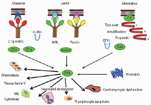
Complement pathways play a critical role in the destruction of invading microorganisms. Working in concert with the adaptive immune system, activation of the three complement pathways (Fig. 81.1) leads to the construction of membrane attack complexes that cause direct lysis and death of the microorganisms (6). During activation, opsonic and chemotactic factors are also generated that facilitate the removal of live and dead organisms from the circulation and from tissues. The importance of these pathways is indicated by the facts that (a) many potentially harmful organisms have mechanisms for avoiding recognition by, or activation of, the complement system, (b) rare complement deficiencies exist in which patients are more vulnerable to infection, and (c) reduced levels of complement proteins in neonates and in the low-protein states of liver disease and nephrotic syndrome may contribute to the susceptibility to infection.

Inherited deficiencies of complement proteins are rare, but affected individuals suffer from an increased risk of bacterial infections. Patients with terminal complement component deficiencies are particularly susceptible to Neisseria sp., including N. meningitidis (7); however, paradoxically, these episodes of infection are generally less severe than in normal populations. It appears that complement activation, while critical for host defense against meningococci, leads to more inflammation and clinical deterioration (8). Although the impact of complement deficiencies on the burden of clinical disease in the pediatric population is negligible, such immune defects serve to demonstrate the complex relationship that exists between susceptibility to, and severity from, an infectious insult.


FIGURE 81.1. The complement and lectin pathways. The lectin pathway of complement is activated by MBL and ficolins. On binding to appropriate targets, MBL-MASP-2 complexes cleave C4 and C2 to form C3 convertase (C4bC2a). MBL-MASP-1 complexes may activate C3 directly. Ficolins also work in combination with the MASPs. The classic and alternative pathways also generate C3 convertase enzymes, which cleave C3. The lytic pathway (C5-C9) is common to all three routes of C3 cleavage. MASP, MBL-associated serine proteases; MASP-1, MBL-associated serine protease-1; MASP-2, MBL-associated serine protease 2. C-INH is C1 esterase inhibitor; CFH is Complement Factor H.

This complex interplay between complement activation, host susceptibility to infection, and inflammatory response is further illustrated by the observation that the complement activation inhibitor Complement Factor H is a susceptibility gene to meningococcal disease (9), but loss of function also predisposes to systemic inflammation (10).


Mannose-Binding Lectin

MBL is a liver-derived plasma protein that recognizes repeating sugar arrays on the surface of many bacteria, fungi, viruses, and parasites. MBL binds to microbes, such as Staphylococcus aureus and N. meningitidis, and activates complement in an antibody- and C1-complex-independent fashion. In contrast to most complement defects, MBL deficiency is common in the population. Three polymorphisms (codons 52, 54, and 57 in exon 1 of MBL-2 gene) are associated with a deficiency of circulating functional MBL. Promoter polymorphisms also contribute to levels of the protein. Approximately one-third of the population is deficient in MBL, with 10% having a profound deficiency.

Children with reduced levels of MBL are at an increased risk for many minor infections of childhood and for more frequent and severe infections in the presence of coexisting immunodeficiencies, such as that caused by chemotherapy for the treatment of cancer. Intensive care is an environment in which many of the most basic defenses against infection are compromised: breaches in skin and airway, poor nutrition, gut hypoperfusion, and acquired “immunoparalysis.” MBL deficiency appears to
be influential in this environment. Children who are admitted to intensive care following infection, trauma, or surgery have a greatly increased risk of developing the systemic inflammatory response syndrome (SIRS) within the first 48 hours of PICU admission if they have MBL deficiency (odds ratio [OR], 7.1; 95% confidence interval [CI], 2.9-19; p = 0.0001). This is true for both infectious (OR, 11; 95% CI, 2-57; p = 0.001) and noninfectious illness (OR, 5; 95% CI, 1.5-19; p

Only gold members can continue reading. Log In or Register to continue

Stay updated, free articles. Join our Telegram channel

Jun 4, 2016 | Posted by in CRITICAL CARE | Comments Off on The Immune System

Full access? Get Clinical Tree

Get Clinical Tree app for offline access