The borders of the Triangle of Petit serve as the major surface anatomic landmarks: the iliac crest inferiorly, the external oblique (EO) muscle anteriorly, and the latissimus dorsi muscle posteriorly.
A line can be drawn along the costal margin cephalad and the iliac crest caudad on the affected side.
Approach and Technique
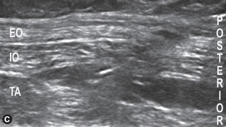
 A high-frequency linear transducer is oriented anterior/posteriorly on the lateral abdominal wall, cephalad to the iliac crest, along the midaxillary line.
The EO, internal oblique (IO), and transversus abdominis (TA) muscles should be visualized.
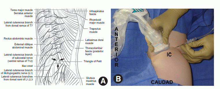
A. Illustration demonstrating the borders of the triangle of Petit and other relevant anatomic landmarks for the TAP block. Figure reproduced from McDonnell JG, et al. RAPM 2007;32:399-404, Fig. 1, pg. 400, with permission. B. The ultrasound transducer is positioned cephalad to the iliac crest, oriented anterior-posterior, and slightly anterior to the mid-axillary line to image the abdominal wall layers in short axis. For preoperative ultrasound-guided TAP catheter insertion, the patient is positioned lateral decubitus, and the needle is inserted posterior to the transducer and directed anteriorly toward the target layer.
US-GUIDED TAP BLOCK
Related posts:


Stay updated, free articles. Join our Telegram channel

Full access? Get Clinical Tree


