Pain refractory to medical therapy
Recurrent pancreatitis secondary to pancreatic duct stenosis
Bile duct stenosis
Gastric outlet obstruction/duodenal obstruction/colonic obstruction
Pancreatic fistula
Pseudocyst
Pancreatic carcinoma
Preoperative Preparation
Preoperative Workup
Just as in all major intraabdominal surgeries, thorough preoperative cardiac risk evaluation should be carried out [5]. In addition, patients with chronic pancreatitis should be screened for ongoing alcohol abuse and referred to a treatment program, if necessary, before undergoing elective resection. In a recent review, 20 % of patients undergoing surgery for chronic pancreatitis retrospectively reported active alcohol abuse at the time of surgery [10].
In cases in which sequelae of chronic pancreatitis have resulted in severe malnutrition (albumin <2), consideration should be given to nutrition supplementation. Enteral feeding is preferable to total parenteral nutrition [9]. Pancreatic enzyme therapy is often required for adequate digestion of food. All patients should receive a bowel regimen such as magnesium citrate the night before surgery to facilitate the creation of a Roux loop, if necessary.
Imaging Studies
Imaging studies are crucial for fully evaluating the extent of disease and for surgical planning. It provides confirmation of chronic pancreatitis, assessment of pancreatic duct diameter, definition of pancreatic anatomy, and the determination of associated disease or malignancy.
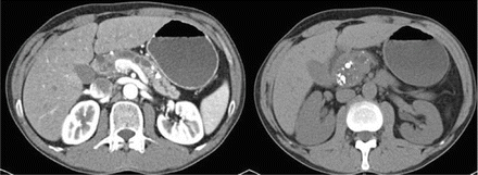
Triple-phase contrast-enhanced, fine-cut, computed tomography is the preferred modality for the evaluation of chronic pancreatitis. The sensitivity of CT scan is almost 100 % for diagnosing advanced disease and readily detects the common sequelae of pancreatitis including the presence of an inflammatory mass, pancreatic pseudocyst, bile duct stricture, pancreatic ductal dilation, and gastric outlet obstruction (Fig. 40.1). In patients with an inflammatory mass, it is crucial to rule out pancreatic adenocarcinoma. If the diagnosis is unclear on axial imaging, additional studies may be required to completely evaluate the patient. Endoscopic retrograde cholangiopancreatogram (ERCP), endoscopic ultrasound (EUS) with biopsy, magnetic resonance imaging (MRI), and magnetic resonance cholangiopancreatogram (MRCP) can provide valuable information in difficult cases. Definitive oncologic resection is indicated if the presence of malignancy is suspected and cannot be ruled out.


Figure 40.1
Preoperative CT scan demonstrating dilated pancreatic duct on contrast scan and severely calcified and diseased pancreatic head on non-contrast images
ERCP is the gold standard for examination of pancreatic ductal anatomy. In chronic pancreatitis, the pancreatic duct displays an irregular contour with multifocal strictures, dilatations, and stones. MRCP provides similar images without the invasiveness of an endoscopy, but is less precise. EUS easily defines pancreatic anatomic relationships and allows for non-invasive tissue sampling under ultrasound guidance [13].
Procedure Selection
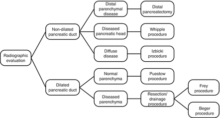
The choice of operation is dependent on pancreatic ductal anatomy and the extent of disease throughout the gland. Operations to palliate abdominal pain either (1) drain a dilated pancreatic ductal system or (2) resect diseased pancreatic parenchyma in cases in which the duct is of normal diameter. The main pancreatic duct normally measures 4–5 mm in the head of the pancreas and gently tapers throughout the body (3–4 mm) and tail (2–3 mm).
Patients with a dilated main pancreatic duct (>7 mm in the body of the gland) are best treated with procedures to decompress and drain the dilated duct (longitudinal pancreaticojejunostomy – Puestow procedure, or longitudinal pancreaticojejunostomy with coring of the head – Frey procedure). On the other hand, patients with a diseased gland and normal pancreatic duct diameter may require resection of the diseased gland. The choice of resection (distal pancreatectomy, pancreaticoduodenectomy, Beger procedure, Izbicki procedure) is dependent on surgeon’s preference and the anatomical extent of disease. Some of these operations (Frey, Beger, Izbicki) have elements of resection and drainage (Fig. 40.2).


Figure 40.2
Treatment algorithm for the surgical management of chronic pancreatitis
Surgical Procedures for Chronic Pancreatitis
Drainage Procedures
Longitudinal pancreaticojejunostomy (Puestow procedure) is performed for the drainage and decompression of a dilated pancreatic duct. Despite providing temporary pain relief, the procedure is associated with approximately 50 % of patients developing recurrent abdominal pain within 5 years. Failure is due to the fact that the procedure may inadequately decompress ducts in the head and uncinate process.
Resection for Chronic Pancreatitis
Patients with parenchymal disease and a normal diameter or narrowed pancreatic duct are candidates for pancreatic resection. The extent of resection is dependent upon the location and extent of disease. Pancreaticoduodenectomy (Whipple resection) may be indicated in patients with an enlarged pancreatic head, containing multiple cysts or calcifications. Distal pancreatectomy is indicated in patients with a normal pancreatic duct and disease confined to the distal gland. However, resection is associated with an increased incidence of postoperative endocrine and exocrine dysfunction.
Combined Resection and Drainage Procedures
Procedures that involve limited pancreatic resection and pancreatic duct drainage attempt to provide permanent pain relief, while avoiding exocrine and endocrine dysfunction. Variations exist based on the method of pancreatic head resection. The Frey procedure combines limited resection by coring of the pancreatic head with unroofing of the dilated pancreatic duct and lateral pancreaticojejunostomy. The Beger procedure is a duodenal-sparing resection of the pancreatic head; drainage is accomplished through a Roux limb to the periampullary pancreas and the remaining tail, either through an end-to-end anastomosis or lateral pancreaticojejunostomy. Both procedures are indicated in severe chronic pancreatitis with an enlarged pancreatic head.
Table 40.2 illustrates the salient differences between the operations employed to treat chronic pancreatitis.
Procedure | Mortality | Endocrine insufficiency | Exocrine insufficiency | Pain relief | Fistula | Comments |
|---|---|---|---|---|---|---|
Puestow procedure | <3 % | Minimal | Unchanged | 85 % | 2 % | Recurrent pain occurs in 40–50 % of patients within 5 years |
Whipple procedure | 40 % | 55 % | 85 % | 7 % | ||
Distal pancreatectomy | 35 % | 30 % | 60 % | 5 % | ||
Frey procedure | <10 %
Stay updated, free articles. Join our Telegram channel
Full access? Get Clinical Tree
 Get Clinical Tree app for offline access

|


