Fig. 9.1
Relative areas of skin excess
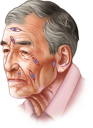
Fig. 9.2
Relaxed skin tension lines can be used to plan excision and primary closure
The surgeon must decide how he will manage margins. In most cases, particularly in areas of excess skin, a well-defined lesion can be primarily excised with comfortable margins. The specimen should, of course be tagged with a stitch and oriented for the pathologist in permanent margins are positive. If a lesion is poorly defined or a complex flap closure is planned, frozen sections can be used to ensure clearance. If these are unavailable, the wound can be safely left open and closed secondarily a week later when permanent section results are available. In the interim, the wound should not be allowed to desiccate.
Once the diagnosis is made and the resection accomplished, adequate closure is then accomplished. Closure may be achieved by direct primary closure, skin grafting, or local flaps. Direct closure is applied in areas of relative skin excess (e.g., cheek, forehead, temple, lip) so as to limit anatomic distortion. Closure should be tensionless particularly near the lower lid and craniad cheek to avoid ectropion. If there is any question, a full thickness graft or flap should be used. Generous undermining (of the scalp or forehead) and even scoring the galea (scalp) can facilitate the process.
Direct closure is accomplished in areas of skin excess (cheek) or specialized areas such as the lip, the helix of the ear, and the eyelid.
In repairing defects, the general principle of “like replaces like” should be applied. For instance, the ideal donor site for a skin graft of the eyelid is the contralateral eyelid (or ipsilateral other lid). Other donor sites are relative areas of skin excess such as pre- or postauricular skin, the temple, and the neck (Fig. 9.3). When skin grafts are used, they should be full thickness grafts. Grafts should be generous, at least the same size as the defect to prevent contracture distortions, particularly around the eyelids (Fig. 9.3). Small grafts on the nose can be taken from the preauricular skin or the lateral forehead, although thick sebaceous nose skin is hard to match. One may consider excision of the whole aesthetic subunit to avoid a noticeable unnatural geographic appearance of small flaps. This should be considered in the nose (Fig. 9.4). A disadvantage of skin grafting, particularly on the nose, is that grafts often do not match the color or texture of the original or surrounding skin.
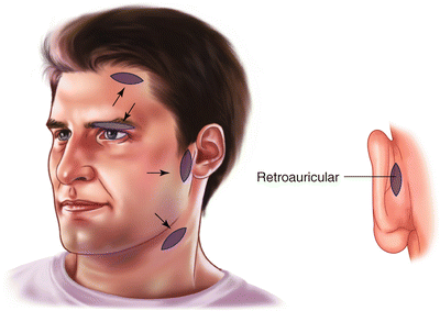
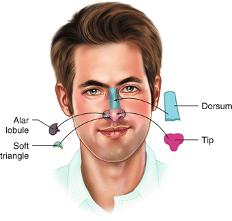
Fig. 9.3
Potential Graft donor sites. An additional site lies behind the ear (retroauricular scalp)
Fig. 9.4
Aesthetic units of the nose
Local flaps have the advantages of color and texture match. This is particularly useful for the nose. They are vascularized so that they can cover full thickness defects straight down to cartilage or bone, allowing for aggressive cancer resection. They are planned to take advantage of relative areas of skin excess. In planning flap closure, one must anticipate the line of maximum tension and make sure that the donor area will close using a “pinch test” (Fig. 9.5). The arc of transposition should be tested or measured to make sure the flap is long enough to close the defect. Because of the generous blood supply of the face and the viscoelastic properties of skin, closure can be performed even under moderate tension as skin creep will allow for eventual capillary refill.
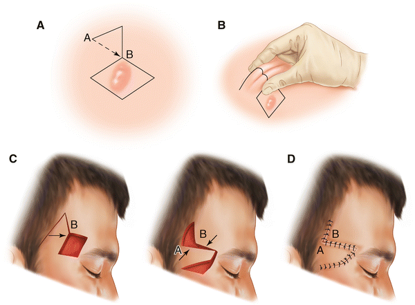
Fig. 9.5
Using a “pinch test” test for adequate closure. A: A rhomboid flap is planned to close an excision of the forehead. Line A–B represents the line of maximum tension. B: The closure is tested with a “pinch test.” C: The flap is rotated to close the defect, while points A and B come together at the point of most tension. D: Final closure
Preoperative Preparation
The patient should be counseled as to the inevitability of facial scars and the possibility of positive margins necessitating further surgery should be discussed.
The patient should be off aspirin for 10 days, if possible, particularly if a graft is anticipated. A sedative may be considered if the patient has anxiety problems or high blood pressure.
The lesion is marked with the patient in the sitting position, taking the RSTL and esthetic subunits into consideration. Loupe magnification is helpful. Having the patient animate the face will accentuate the RSTL and help orient the resection. A pinch test is used when primary closure or flap closure is anticipated (across the maximum line of tension). Mark all anatomic lines to be approximated before injection (e.g., the vermillion, the alar rim, the helix of the ear, etc). Donor sites for grafts are marked.
Operative Strategy
Surgical management of facial neoplasia must focus on complete excision of the carcinoma. That is to say that excision must be perpendicular through all layers of the skin, and deeper to perichondrium or bone if necessary. Once resection is adequate, then closure is planned. One should not allow the anticipated method of closure to dictate the limits of resection, as it is the resection that must be complete. There are no magic numbers for margins. Margins can be planned so as to allow for direct closure. If adequate margins cannot afford direct closure, grafts or flaps should be used. Occasionally, excision of an entire aesthetic unit may give a better result after subsequent grafting than small, patchwork grafts. This is particularly true of the nose. All specimens are tagged with a stitch, the specimen for the pathologist. Make sure all margins are clear before closing with a flap, so as not to bury and obscure a recurrence.
Related posts:


Stay updated, free articles. Join our Telegram channel

Full access? Get Clinical Tree


