Tumor thickness
Recommended clinical margins
In situ
0.5 cm
≤1.0 mm
1.0 cm (category 1)
1.01–2 mm
1–2 cm (category 1)
2.01–4 mm
2.0 cm (category 1)
>4 mm
2.0 cm (category 1)
Table 11.2
Indications for sentinel lymph node biopsy (clinically node negative patients)
Discuss and consider: |
Breslow depth ≥0.76 mm, no ulceration, no ulceration, mitotic rate <1 per mm2 |
Discuss and offer: |
Breslow depth ≥0.76 mm, and any of the following: |
Ulceration |
Mitotic rate ≥1 per mm2 |
Lymphovascular invasion |
Breslow depth >1 mm (any) |
Preoperative Preparation
Most lesions can be excised using an elliptical, longitudinal excision of the scar and tumor bed following Langer’s lines on the trunk and proximal extremities (Fig. 11.1). The Breslow depth is used to determine the margins of the resection. In situ lesions are routinely resected with margins of 0.5–1.0 cm. T1 melanomas, with a Breslow depth of less than or equal to 1.0 mm, are excised with 1 cm margins. T2 lesions, 1.01–2 mm in depth can be safely excised in most locations with a 1–2 cm margins. An exception may be the scalp, where consideration should be given to perform a 2 cm wide local excision, due to the higher rate of local recurrences in this location. T3 lesions (2.01–4 mm in depth) should be excised with 2 cm margins. Thick, T4 (>4.0 mm) lesions are also excised with a 2.0 cm margin.


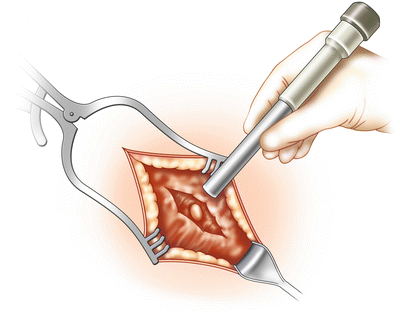
Fig. 11.1
Identification of a sentinel lymph node with a sterile gamma probe
Thin melanomas with a Breslow’s depth of ≤0.75 mm (and especially those with no ulceration and no mitotic figures) have a low likelihood to metastasize to regional lymph nodes (<5 %). Patients with lesions ≤0.75 mm thick are usually not offered a sentinel lymph node biopsy, unless they have a lesion with one or more adverse prognostic indicators. Such factors include ulceration, lymphovascular invasion, a high mitotic count, or Clark’s level >III (if the mitotic count cannot be determined). All patients with a measured Breslow thickness ≥0.76 mm, with clinically negative nodes (clinical Stage I and II), are appropriate candidates for sentinel node biopsy. Almost all procedures are performed an outpatient basis. We prefer a single dose of a first generation cephalosporin, or clindamycin in the setting of a penicillin allergy, as prophylaxis. As per current Surgical Care Improvement Project (SCIP) guidelines, the perioperative antibiotics should be administered prior to creation of the incision.
Operative Strategy
Lymphoscintigraphy should be performed in every case in which sentinel lymph node biopsy is performed. Preoperative lymphoscintigraphy helps identify the number and relative location of the sentinel lymph node(s) in a particular basin. The mapping may also identify in transit sentinel nodes that may be a source of regional or distant metastasis if not recognized and excised. The lymphoscintigraphy is typically done on the morning of surgery, 1–2 h prior to the excision.
Operative Technique
The patient is seen in nuclear medicine the morning of the surgical excision. A four-point intradermal injection technique is used to inject 0.5–1 mCi of 99m-technetium sulfur colloid into the lesion. Early and frequent images using a gamma camera are then obtained in anterior and lateral views in order to identify the sentinel lymph nodes to be biopsied. At surgery, just prior to prepping and positioning the patient, 1–3 mL of 1 % isosulfan blue dye (Lymphazurin) is injected in a sterile fashion in the area around the lesion or biopsy scar. 1 cc of 1 % plain lidocaine may be added to the lymphazurin for patient comfort.
The patient is prepped and draped in the usual sterile fashion including the nodal basins from which the sentinel nodes will be harvested. The patient should be positioned to allow for nodal biopsy along with excision of the malignant lesion if at all possible. The patient can always be repositioned if needed between the sentinel node biopsy and the wide local excision. A time out is performed with the side and site of the surgery verified.
Stay updated, free articles. Join our Telegram channel

Full access? Get Clinical Tree


