 Pt’s generally seek help only if their sunburn is severe. There will be a history of extended exposure to sunlight or to an artificial source of ultraviolet radiation, such as a sunlamp. Inquire as to whether or not the pt is using a photosensitizing drug (e.g., tetracyclines, thiazides, sulfonamides, phenothiazines). Sunburn prevalence increased among US adults from 1999 to 2004 from 31.8% to 33.7% (MMWR 2007;56:524-528).
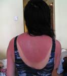
S/s: the skin becomes red, painful, and abnormally warm after sun exposure. There may be systemic complaints that include nausea, chills, and fever. The affected areas are erythematous and are accompanied by mild edema. The more severe the burn, the earlier it will appear and the more likely it will progress to edema and blistering. Onset of sx’s is usually delayed for 2-4 hours. Maximum discomfort usually occurs after 14-20 hours, and sx’s last between 24 and 72 hours. 58% of those age 18-29 report having a sunburn in the past year (Am J Prev Med 2002;23:91-97). Higher income, binge drinking, and student status are all independent risk factors for sunburn in the US (Arch Derm. 2003;139:1003–6).
Tx: Cool / moist compresses: Take a cool shower or bath (avoid bath salts, oils, and perfumes to prevent sensitivity reactions). Immerse the sunburned area in cool water or apply cool / moist compresses: water or Burrow’s solution (Domeboro Powder Packets-1 packet in 1 pint of water) as often as desired to relieve pain. Apply moisturizing lotion to affected areas. Calamine or Aloe Vera gel (keep them in the refrigerator). Avoid further sun exposure until the burn has resolved. Drinking plenty of water helps to replace fluid losses.
Do not: use salve, butter, or ointment. Do not break blisters.
Topical steroid: Rx a potent one such as Ultravate (Halobetasol) 0.05% BID for 3 days if begun within 48hr of exposure or use a topical steroid spray such as dexamethasone (Decaspray) and using an emollient such as Lubriderm. A low-dose OTC (0.5%-1%) hydrocortisone cream can be helpful in reducing the burning sensation and swelling and speeding up healing (Clin Exp Dermatol. 2002;27:314-8). A RCT with 20 pt’s concluded that a topical moderate or high-potnecy corticosteroid did not provide a clinically useful decrease in acute sunburn reaction when applied 6 or 23 hours after UV exposure (Arch Derm 2008;144:620-24).
Prednisone 40-60mg qd x 3d or Medrol dose pack or IM Aristocort if severe as this will reduce inflammation, swelling, pain, and itching. Analgesics (ASA or other NSAIDs). There may be an incr risk of scar formation in those taking NSAIDs (J Am Acad Derm 2001;45:746).
Pearls: Inquire as to whether or not the pt is using a photosensitizing drug (e.g., tetracycline’s, thiazides, sulfonamides, phenothiazines) and have the pt discontinue its use. Do not allow the pt to use OTC sunburn medications that contain local anesthetics (benzocaine, dibucaine or lidocaine). They are usually ineffective or only provide very transient relief. In addition there is the potential hazard of sensitizing the pt to these ingredients. If pt has significant areas of débrided skin, Rx Silvadene cream or Polysporin ointment, Bactroban ointment to prevent secondary infection.
ICD-9 Code: 692.71 Sunburn
Compounded Rx – Insect Bite/Sunburn Gel: Hydrocortisone 1 g + Pramoxine HCl 1 g + Diphenhydramine HCl 2 g + Menthol 300 mg + Compounded Rx – Wound Care Mixture: Phenol 200 mg + Zinc oxide 12 g + 70% Ethanol + Calcium hydroxide solution aa qs 100 mL. Accurately weigh/measure each ingredient. Prepare 100 mL of the vehicle using equal parts of 70% ethanol and calcium hydroxide solution (lime water). Dissolve the phenol in about 75 mL of this vehicle. Sprinkle the zinc oxide powder on the phenol-vehicle mixture. Add additional vehicle to volume and mix well. Package and label.
Children: Many studies have suggested that childhood sun exposure is a significant risk for later development of skin cancers. In addition, sun exposure to unprotected eyes is also a major risk for later cataracts. Given that up to 80% of lifetime UV exposure occurs before age 18 years, sun protection in childhood is of paramount importance. It is strongly advised that parents shield children younger than 6 months from any excess sun with hats, clothing, umbrellas, and shade. In older age groups, children should apply sunscreens heavily 30 minutes before exposure. They should use sunscreens with a SPF of 15 or greater. If a person normally burns in 1 hour, an SPF of 15 will protect them from a burn for 15 hours. Similarly, people who burn in 10 minutes will only obtain 150 minutes of protection with the same sunscreen. Children with light eyes and lighter complexions would do better with an SPF of 30. Reapplication after water immersion or sweating is also advocated. Still, a sunscreen’s protection is not perfect, and hats, long sleeves, and pants are extremely helpful, especially during the peak hours of 11 AM to 2 PM, when children should avoid sun exposure. Several manufacturers make clothing with exceptionally high SPFs. Providers should make parents aware that children need these sun protection measures on cloudy days and especially when near reflective surfaces, such as water and snow.
**Ref: (Trauma, 4th ed., 2000, McGraw-Hill) (Ambulatory management of burns. Am Fam Phys 2000;62:9) (Electrical injury, Curr Prob Surg 1997:34:677) (Environmental Emergencies, Chpt 74, 1996) (Thermal injury. Crit Care Clin. 1999;15:333-52) (Current status of burn resuscitation. Clin Plast Surg. 2000;27:1-10) (Hydrofluoric acid: a review of toxicity. J Emerg Med. 1992;10:163-8) (Caustic ingestion injuries. Gastroenterol Clin North Am. 1991 Dec;20:847-57) (Chemical burns of the upper extremity. Hand Clin. 1990;6:253-9) (Decontamination and management of hazardous materials exposure victims. Ann Emerg Med. 1994;23:761-70) (Electrical burns. Clin Plast Surg. 2000;27:133-43)
(Electrical and lightning injuries. Crit Care Clin. 1999;15:319-31) (Electric injury, part I: tx priorities, subtle diagnostic factors, and burns. J Emerg Med. 1999;17:977-83) (Electric injury, Part II: specific injuries. J Emerg Med 2000;18:27) (Electric injury, Part III: cardiac monitoring indications, the pregnant pt and lightning. J Emerg Med 2000;18:181) (Electrical injuries. So J Med 2000;93:12) (Recommendations for lightning safety. JAMA 1999;282:1132-1133) (Electrical injuries. J Crit Illness 2002;17:3) (ABC of burns. BMJ 2004;328:1427-1429)
Links: Intro & PP | Risks | PV | Initial Hx & Exam | Stroke Scales & Codes | Initial W/u – Eval | Stroke Syndromes (location) | Sensory Exam | Ischemic (Infarction) / Embolic | Lacunar | Hemorrhagic (Subdural / Epidural) | Subarachnoid Hemorrhage & Intracranial aneurysm | PV & Long-term Tx | Recovery | Transient Ischemic Attack (TIA) & TNA | Cerebral Circulation Diagram | Carotids | Hematoma | Thrombolytics | PFO | Young (<45yo) Pt’s | Imaging Studies | Silent |
ICD-9: CVA, acute 436. TIA 435.9.
ICD-10 codes:
(I60.0) Subarachnoid hemorrhage from carotid siphon and bifurcation
(I60.1) Subarachnoid hemorrhage from middle cerebral artery
(I60.2) Subarachnoid hemorrhage from anterior communicating artery
(I60.3) Subarachnoid hemorrhage from posterior communicating artery
(I60.4) Subarachnoid hemorrhage from basilar artery
(I60.5) Subarachnoid hemorrhage from vertebral artery
(I60.6) Subarachnoid hemorrhage from other intracranial arteries
(I60.7) Subarachnoid hemorrhage from intracranial artery, unspecified
(I61) Intracerebral hemorrhage
(I61.0) Intracerebral hemorrhage in hemisphere, subcortical
(I61.1) Intracerebral hemorrhage in hemisphere, cortical
(I61.2) Intracerebral hemorrhage in hemisphere, unspecified
(I61.3) Intracerebral hemorrhage in brain stem
(I61.4) Intracerebral hemorrhage in cerebellum
(I61.5) Intracerebral hemorrhage, intraventricular
(I61.6) Intracerebral hemorrhage, multiple localized
(I62) Other nontraumatic intracranial hemorrhage
(I62.0) Subdural hemorrhage (acute)(nontraumatic)
(I62.1) Nontraumatic extradural hemorrhage
Nontraumatic epidural hemorrhage
(I63) Cerebral infarction
(I63.0) Cerebral infarction due to thrombosis of precerebral arteries
(I63.1) Cerebral infarction due to embolism of precerebral arteries
(I63.2) Cerebral infarction due to unspecified occlusion or stenosis of precerebral arteries
(I63.3) Cerebral infarction due to thrombosis of cerebral arteries
(I63.4) Cerebral infarction due to embolism of cerebral arteries
(I63.5) Cerebral infarction due to unspecified occlusion or stenosis of cerebral arteries
(I63.6) Cerebral infarction due to cerebral venous thrombosis, nonpyogenic
(I64) Stroke, not specified as hemorrhage or infarction
(I65) Occlusion and stenosis of precerebral arteries, not resulting in cerebral infarction
(I65.0) Occlusion and stenosis of vertebral artery
(I65.1) Occlusion and stenosis of basilar artery
(I65.2) Occlusion and stenosis of carotid artery
(I65.3) Occlusion and stenosis of multiple and bilateral precerebral arteries
(I65.8) Occlusion and stenosis of other precerebral artery
(I65.9) Occlusion and stenosis of unspecified precerebral artery
(I66) Occlusion and stenosis of cerebral arteries, not resulting in cerebral infarction
(I66.0) Occlusion and stenosis of middle cerebral artery
(I66.1) Occlusion and stenosis of anterior cerebral artery
(I66.2) Occlusion and stenosis of posterior cerebral artery
(I66.3) Occlusion and stenosis of cerebellar arteries
(I66.4) Occlusion and stenosis of multiple and bilateral cerebral arteries
(I66.5) Occlusion and stenosis of other cerebral artery
(I66.6) Occlusion and stenosis of unspecified cerebral artery
(I67) Other cerebrovascular diseases
(I67.1) Cerebral aneurysm, nonruptured
(I67.2) Cerebral atherosclerosis
(I67.3) Progressive vascular leukoencephalopathy
Binswanger’s disease
(I67.4) Hypertensive encephalopathy
(I67.5) Moyamoya disease
(I67.6) Nonpyogenic thrombosis of intracranial venous system
(I67.7) Cerebral arteritis, not elsewhere classified
(I68) Cerebrovascular disorders in diseases classified elsewhere
(I69) Sequelae of cerebrovascular disease
 Intro: Stroke is an interruption of normal cerebral blood flow causing neurological deficits. A stroke is focal brain dysfunction due to ischemia that may arise from atherosclerotic narrowing of a blood vessel, an embolus, hemorrhage, or other causes. It is an acute neurologic d/o produced by nontraumatic injury in the central nervous system that is vascular in origin, it is accompanied by focal rather than global neurologic dysfunction that persists for >24 hours or results in death within the first 24 hours. Up to 15% turn out to be a TIA. ~75% of the strokes are ischemic (thrombotic, embolic, hypoperfusion), 25% hemorrhagic (intracerebral or subarachnoid). The 3rd leading cause of death and the leading cause of long-term disability in the USA–> 25% die, 15% risk re-stroke in next month w/o tx, ~30% are permanently disabled with ~20% requiring institutionalized care. The leading cause of disability in the USA. H-A is most common sx, MS change, focal deficit. Need to clear C-spine if falls. 50% have HTN (the most important risk factor for most strokes), 30% smoke, 40% with incr chol. At least 20% of strokes have no known cause and are labeled as cryptogenic.
• Women tend to have more nontraditional sx’s such as pain, change in level of consciousness or disorientation (Ann Emerg Med 2002;40:461-3), compared to the usual hemiparesis or imbalance in males.
• The lifetime risk for stroke in middle-aged adults at about 1 in 6 (18%) based on Framingham data of pt’s followed for 51 years (hypertensive participants had a consistently greater risk for stroke than did normotensive participants) (Stroke 2006;37:345-50).
• Data from the Oxford Vascular Study with 91,106 pt’s suggest that cerebrovascular events are more common than coronary events, despite a significant decline in stroke rates during the past 20 years and a recent increase in MI rates (following use of more-sensitive diagnostic criteria) (Lancet 2005;366:1773-83)…..more than half of all acute vascular events occurred among people age 75 or older.
• Ischemic stroke pt’s who arrive at the hospital in an ambulance (52%) are more likely to meet the deadline for the use of tPA- 3 hours from sx onset (MMWR 2007;56;474-478) (median wait for imaging was 44 minutes)….need educating the public regarding the s/s of stroke and the importance of telephoning for ambulance transport.
• About 70% of patients who suffer a minor stroke or TIA do not correctly recognize their symptoms, and 30% delay seeking medical attention for more than 24 hours a study on 1000 consecutive patients (459 presented after TIA and 541 after minor stroke) concludes (Stroke. Published online April 15, 2010)…..Of patients with TIA, 208 (47%) sought medical attention within 3 hours, the window of time during which thrombolytic therapy is optimally administered, as did 234 (46%) minor stroke patients; 300 (67%) TIA patients and 400 (74%) of those with minor stroke sought medical attention within 24 hours……more likely to delay if they had no motor and speech symptoms (P < .001), the event lasted less than 60 minutes (P < .001), or they were younger than 60 years (P = .075).
PP: Cerebral cortex: neuron cell bodies (gray matter). Immediately below is the centrum semiovale with myelinated axons (white matter). Surrounding the axons is supporting tissue (glia). Normal cerebral blood flow is ~50-60 ml/100g of brain tissue/min. If flow <20-40 ml/100g / min then get neuronal dysfunction. If <10-15 ml/100g/min then irreversible tissue damage occurs. A sublethal noxious stimulus is known to protect the brain from subsequent ischemic insult (J Neurosci 2007;27:7083-7093), this “preconditioning” is mediated by changes in levels of nitric oxide and reactive oxygen species. See Stroke Syndromes (location) | Ischemic (Infarction) / Embolic |
Risk: Stroke. 2006;37:online May 4, 2006: Age (risk doubles with each successive decade after 55 years). Men have higher age-specific stroke incidence rates than women, except for those aged 35 to 44 years and older than 85 years, groups in which women have higher rates. African Americans and some Hispanic Americans have higher stroke rates and stroke mortality vs European Americans. OC use and pregnancy contribute to stroke risk in women. The odds of stroke are more than doubled in individuals older than 50 years who had low birth weights of <2000 g vs those with birth weights >4000 g. FHx. The presence of atherosclerotic disease overall confers increased stroke risk. Ischemic stroke risk doubles for cigarette smokers, and hemorrhagic stroke risk increases 2 to 4 times vs nonsmokers. Sickle cell disease. Carotid stenosis. Sleep apnea. Passive smoking. Obesity (Arch Int Med 2002;162:2557-62). Afib + rheumatic heart dz (18X), prior TIA/stroke (10X), HTN (6X), Afib (6X), CHF (5X), angina/AMI (3X), DM (3X), heavy ETOH use (3X risk, ?60g/d), smoking (3X), valvular dz. Protease inhibitors linked to increased stroke risk. In pt’s with CAD, serum total homocysteine level is directly related to the risk of ischemic stroke, if >17.4 micromols/L were 4.6X risk than if <11.4 (Stroke 2003;34:423).
• CRP may predict risk of stroke independent of other risk factors (Circulation 2003;107:4575). If FHx of stroke, then more than 2X as likely to experience an ischemic stroke (Stroke 2003;34:4220).
• Respiratory infections may increase the risk of large vessel and cardioembolic stokes (infection alters platelet aggregation and coagulation), not ischemic (Stroke 2003;34:452-57).
• HTN (4-fold risk) and low levels of von Willebrand factor are independent and synergistic risk factors for intracranial hemorrhage (Stroke 2004;35:826-830), pt’s in the lowest vWF tertile were 73% more likely to have an ICH than those in the highest tertile, with HTN there was a 9-fold risk. Infection with H pylori bearing cytotoxin-associated gene-A (CagA) appears to be associated with ischemic stroke and TIA’s (Stroke 2004;35:1800-1804). Individuals with serum Ab’s to periodontal pathogens are more likely to experience stroke than are their seronegative counterparts (Stroke 2004;35:2020-2023,2029-2035), also, tooth loss and long-term periodontitis are related to carotid artery plaque in men.
• Chemotherapy appears to increase the risk (2-fold) of stroke in pt’s with breast cancer regardless of tamoxifen use (J Natl Cancer Inst 2004;96:1528-1536).
• Findings from a genotype-phenotype analysis suggest that high homocysteine levels may play a causal role in stroke (Lancet 2005;365:194-195,224-232). There appears to be an association between migraine with aura and other headaches with aura, and an increased risk of stroke and TIA (Neurology 2005;64:1573-1577) (OR 2.81-5.46).
• Moderate alcohol use (1 to 6 drinks per week, U-shaped relationship) reduces the risk of ischemic stroke, except among individuals who are positive for apolipoprotein E4 (Stroke 2005;36:1830-1836). Both preeclampsia and gestational diabetes put women at increased risk for stroke in later years (odds ratios of 2.06 and 2.44, respectively) (Am Neurol Assoc. Abstract 222. Presented Sept 27, 2005)…..strokes occurred an average of 13.5 years after delivery, and the women were an average of 40.1 years old. Retinopathy, as determined with retinal photography, is an independent predictor of stroke or stroke-related death in older adults without diabetes (Neurology 2005;65:1005-1009)….similar anatomic characteristics and other characteristics with the blood vessels in the brain. A pronounced increase in the risk of stroke is seen following an MI (Ann Intern Med 2005;143:785-792) (44-times higher than the rate in the first 30 days, 2.5-fold in the next 3-years). Increases in particulate air pollution raise the risk of admission for ischemic stroke, but not hemorrhagic stroke (Stroke 2005;36;Oct).
• People who smoked heavily in the past before quitting continue to carry a long-term risk for stroke based on atherosclerotic burden (American Stroke Association’s International Stroke Conference 2006;Feb 24).
• The presence of renal dysfunction, even of mild severity (GFR <60), seems to raise the risk of ischemic stroke or TIAs by 54% in pt’s with cardiovascular disease (Neurology 2006;67:224-228). Missing teeth, loss of bone around teeth and reduced periodontal health appear to be associated with an increased risk of ischemic stroke (J Periodontal 2006;77:1744-1754). LV systolic dysfunction, even mild, is associated with an increased risk of ischemic stroke (Stroke 2006;37:1715-1719) (OR for any degree of LVD was 3.92, for moderate/severe LVD and mild LVD, the ORs were 3.88 and 3.96, respectively)….one possibility is that LVD promotes increased blood stasis in both the LV and left atrium, increasing the chance of thrombus formation and the risk of embolic stroke. Individuals with a sibling who has had a stroke have an increased risk of sustaining a severe stroke and greater neurologic deficits (Neurology 2006;67:1396-1402).
• A study has shown a relationship between the risk for fatal stroke and fine and ultrafine particulate (< 0.1 µm) air pollution (Stroke. Published online February 15, 2007) (elderly people should avoid spending unnecessary time in traffic, whether it is in a vehicle or walking, especially if they suffer from cardiovascular diseases)(only significant during the warm season).
• A genetic defect (H63D and C282Y single neucleotide polymorphisms [SNPs]) that causes hereditary hemochromatosis (affecting at least 25% of the European population) predicts a significantly increased risk (2 to 3-fold) for ischemic cerebrovascular disease (ICVD) and ischemic stroke (Neurology. 2007;68:1025-1031)…….have previously been linked to Alzheimer’s disease, Parkinson’s disease, amyotrophic lateral sclerosis, multiple sclerosis, cerebrovascular disease (carotid atherosclerosis), and stroke….mechanism unclear as the data suggest plaque buildup in the arteries and iron overload are not to blame.
• Serum levels of complement factors C3 and C4 were predictors of stroke risk over two years in a cohort of men referred for coronary angiography, with C4 emerging as predictive independent of other inflammation markers (Am J Cardiol 2007;100:164-168). Low vitamin B12 plasma levels, especially in combination with low folate levels, are associated with an increased risk of ischemic stroke or TIA (RR = 2.24)(Stroke 2007;38:2912-2918).
• Poor kidney function, which is known to increase cardiovascular risk, might also be associated with subclinical markers of cerebral small vessel disease, independent of cardiovascular risk factors (Stroke. 2008;39:55-61).
• Following an acute coronary syndrome, the risk of stroke is up to 10 times higher (2% to 3%) than the 1-month risk of stroke among patients with stable angina or remote myocardial infarction (Arterioscler Thromb Vasc Biol 2008;28:142-147)….related to the intensity of the inflammation following a cardiac event and the extent of heart damage…..intensive cholesterol-lowering with a high-dose statin soon after a heart attack not only prevents repeat heart problems, but it also prevents stroke, probably by having similar effects on dampening the inflammation in arteries that supply the brain as well as those that supply the heart. Slow walking speed was found to be a strong predictor of an increased risk for incident ischemic stroke among postmenopausal women independent of other established risk factors for stroke (Stroke. Published online February 21, 2008)…..Reduced walking speed has previously been associated with an increased risk for falls, functional disability, hospitalizations, dementia, white matter hyperintensities and lacunar infarcts, and incident stroke among older adults.
• Unintentional regular daytime dozing can more than quadruple stroke risk in elderly patients and significantly increase their risk (4.5-fold vs counterparts who did not doze off during the day) for other vascular events (American Stroke Association International Stroke Conference 2008: Abstract 94. Presented February 21, 2008)…..poor or diminished quality of sleep to an increased risk for vascular events.
• Full-blown panic attack are associated with subsequent cardiovascular or cerebrovascular events (Arch Gen Psychiatry 2007;64:1153)…unkown if the mechanism ischemic or neurogenic….possibly related to takotsubo-like cardiomyopathy affects seen mainly postmenopausal women. A meta-analysis of data from 10 published studies involving 140,231 participants and 3,266 strokes found that participants with proteinuria had a 71% greater risk of stroke compared with those without proteinuria (Am J Kidney Dis 2009;53:417-425)…..support the hypothesis that proteinuria is an independent risk factor for stroke.
• Parental stroke before the age of 65 years is associated with a 3-fold increased risk for stroke in offspring, according to data from the Framingham Heart Study (Circulation. 2010;121:1304-1312)…..“Although there are many environmental determinants of stroke, such as smoking, physical inactivity, and diet, we also know that various factors are under genetic as well as environmental control, such as hypertension, diabetes, high cholesterol, and obesity.
• Coronary artery calcification (CAC) independently predicts future stroke risk in people considered to be at low and intermediate risk, according to a study (STROKEAHA.111.678078 Published online before print February 28, 2013)…..In a multivariable Cox regression, log10(CAC+1) was an independent stroke predictor (hazards ratio, 1.52 [95% confidence interval, 1.19–1.92]; P=0.001) in addition to age (1.35 per 5 years [1.15–1.59]; P<0.001), systolic blood pressure (1.25 per 10 mm Hg [1.14–1.37]; P<0.001), and smoking (1.75 [1.07–2.87]; P=0.025).
• Patients with cancer who have none of the conventional stroke risk factors are at increased risk for stroke due to cancer-related hypercoagulation (Stroke. 2012;43:3029-3034)…..Unidentified stroke etiology was significantly more frequent in the cancer group (48%) than in controls (27%) (P < .001)….The prevalence of deep-vein thrombosis and pulmonary embolism was similarly significantly higher in patients with cancer than in controls: 8% vs 1% (P < .01). “Metastatic disease itself was significantly more frequent in patients with cancer with unidentified and/or stroke-associated stroke etiology (59%) than in patients with cancer with conventional stroke etiology (28%; P < 0.05),” the investigators add.
Diet: Women who consume at least 102 g red meat a day have a 42% higher risk for cerebral infarction than those who eat 25 g or less red meat daily (Stroke. Published online December 16, 2010)……At least 1 previous study has shown that the risk for hypertension, which is implicated in stroke, is higher among meat eaters……Red meat contains saturated fat and cholesterol, which have also been linked to poor cardiovascular health…..Red meat is also rich in hemo iron — a nutrient known to catalyze the formation of hydroxyl radicals that are powerful prooxidants.
• A study again has found that higher consumption of both processed and unprocessed red meat is associated with a higher risk for stroke, whereas intake of poultry was associated with a reduced risk according to data from both the Nurses’ Health Study (NHS) and the Health Professionals Follow-Up Study (HFPS)(Stroke. Published online December 29, 2011)……with processed red meat (per 1 serving per day) RR= 1.32….for unprocessed red meat (per 1 serving per day) RR= 1.16……Poultry consumption RR = 0.77 (95% confidence interval, 0.62 – 0.95; P = .02)……These associations were independent of other major protein sources, fruits and vegetables, and other stroke risk factors….The findings were similar for ischemic and hemorrhagic stroke……..by substituting 1 serving of red meat with a serving of nuts or fish, the stroke risk was reduced by 17%, and by replacing the red meat with low-fat dairy, the risk went down by 11%. Substituting 1 serving of red meat with a serving of poultry resulted in a 27% reduction in stroke risk.
Depression: Higher levels of depressive symptoms are associated with subclinical cerebrovascular disease (based on MRI) in older women but not men (Am J Geriatr Psychiatry 2010;March 11th online).
• Psychological distress (i.e., depressive symptoms), but not major depression, increases the risk for stroke according a analysis of 595 pt’s (Neurology 2008;70:788)….stroke risk increased by 11% with each standard-deviation increase in psychological distress. A study suggests an association between depression and an increased risk (18%) for first-ever stroke in elderly subjects (HR 2.7)(Stroke 2008;39:1960-1965).
• Sx’s of depression appear to increase the risk of stroke in men with risk factors for CAD (Stroke 2004;36). An analysis from the UK European Prospective Investigation into Cancer (EPIC)–Norfolk population-based study has shown a relationship between psychological distress and increased risk for stroke (Neurology. 2008;70:788-794)….No such link was seen between episodic major depressive disorder and stroke in this analysis……incidence of depression after stroke was 34% vs a rate of 13% among age-matched controls.
• The risk of developing dementia is high after stroke (21.5% at 2yrs), the characteristics of post-stroke dementia appear to shift from an Alzheimer disease-type picture in the first years after stroke to a vascular dementia type in later years (Neurology 2004;62:2193-2197).
• A systematic review and meta-analysis finds that depression increases the risk for stroke (JAMA 2011;306:1241)…. those with depression had a 45% higher risk for stroke, a 55% higher risk for fatal stroke (evaluated in 8 studies), and a 25% higher risk for ischemic stroke (evaluated in 6 studies)……depression could lead to stroke via several mechanisms, including prothrombotic and proinflammatory effects, but more studies are needed to clarify whether depression is causal or simply a marker for stroke.
Risk factors for ischemic and intracerebral hemorrhagic stroke in 22 countries:
(the INTERSTROKE study. Lancet 2010; DOI:10.1016/S0140-6736(10)60834-3):
* 90% of population-attributable risks were attributable to 10 risk factors.
* Hypertension identified by self-report increased the risk for all stroke (odds ratio [OR], 2.64), ischemic stroke (OR, 2.37), and intracerebral hemorrhagic stroke (OR, 3.80).
* Hypertension based on self-report or blood pressure of more than 160/90 mm Hg increased the risk for all stroke (OR, 3.89), ischemic stroke (OR, 3.14), and intracerebral hemorrhagic stroke (OR, 9.18).
* Current smoker, defined as any tobacco use in the prior 12 months, vs never-smoker or former smoker increased the risk for all stroke (OR, 2.09), ischemic stroke (OR, 2.32), and intracerebral hemorrhagic stroke (OR, 1.45).
* The highest vs the lowest tertile of waist-to-hip ratio had greater risk for all stroke (OR, 1.65), ischemic stroke (OR, 1.69), and intracerebral hemorrhagic stroke (OR, 1.41).
* High diet risk score indicating an unhealthy cardiovascular diet was linked with an increased risk for all stroke (OR, 1.35), ischemic stroke (OR, 1.34), and intracerebral hemorrhagic stroke (OR, 1.41).
* Self-reported diabetes mellitus had an increased risk for all stroke (OR, 1.36) and ischemic stroke (OR, 1.60), but not intracerebral hemorrhagic stroke.
* More than 30 alcoholic drinks per month or binge drinking vs never or former alcohol use was linked with an increased risk for all stroke (OR, 1.51), ischemic stroke (OR, 1.41), and intracerebral hemorrhagic stroke (OR, 2.01).
* Alcohol intake of 1 to 30 drinks per month was linked with a decreased risk for ischemic stroke (OR, 0.79).
* Psychosocial stress was linked with an increased risk for all stroke (OR, 1.30), ischemic stroke (OR, 1.30), and intracerebral hemorrhagic stroke (OR, 1.23).
* Depression was linked with an increased risk for all stroke (OR, 1.35) and ischemic stroke (OR, 1.47), but not intracerebral hemorrhagic stroke.
* Cardiac causes were associated with an increased risk for all stroke (OR, 2.38) and ischemic stroke (OR, 2.74), but not intracerebral hemorrhagic stroke.
* Higher ratio of apolipoprotein B to apolipoprotein A1 was linked with an increased risk for all stroke (OR, 1.89) and ischemic stroke (2.40), but not intracerebral hemorrhagic stroke.
* Regular physical activity was linked with a lower risk for all stroke (OR, 0.69) and ischemic stroke (OR, 2.40), but not intracerebral hemorrhagic stroke.
* Ratio of non-high-density lipoprotein to high-density lipoprotein cholesterol was linked with an increased risk for ischemic stroke and a decreased risk for intracerebral hemorrhagic stroke.
* Body mass index was not linked with stroke risk.
• Aspirin resistance is common in patients with acute ischemic stroke and is associated with more severe stroke and larger infarct size, according to a study (Arch Neurol. Published online November 19, 2012)…..researchers tested for aspirin resistance among 90 patients with acute ischemic stroke who had been receiving aspirin therapy (100 mg daily) for an average of 5 years before stroke. Of these, 26 (28.9%) were aspirin resistant, defined by more than 550 aspirin reaction units (ARU) on the rapid platelet function assay VerifyNow (Accumetrics). Every 1-point increase in ARU was associated with a statistically significant 0.03-point increase in NIHSS score (95% CI, 0.01 – 0.04; P = .001). This corresponded to an approximate median increase of 1 point in NIHSS score for every 33-point increase in ARU. NOTE: Taking ASA BID seems to mitigate the risk in 75%.
SSRI’s: Although antidepressants were not associated with coronary heart disease risk, SSRIs were associated with higher stroke risk and both SSRIs and TCAs with higher all-cause mortality according to data from the Women’s Health Initiative (Arch Intern Med 2009;169:2128)…..Compared with no antidepressant use, SSRIs were associated with higher risk for stroke (hazard ratio, 1.4), primarily hemorrhagic (HR, 2.1), and with all-cause mortality (HR, 1.3). TCAs also were associated with higher all-cause mortality (HR, 1.7). Absolute event numbers were low: Annualized stroke rates per 1000 person-years were 3.0 in nonusers and 4.2 in SSRI users, and death rates were 7.8 (nonusers), 12.8 (SSRI users), and 14.1 (TCA users) per 1000 person-years.
The left atrial septal pouch (LASP), that could be an important, hitherto-unrecognized source of thrombus with ischemic stroke or cryptogenic stroke (JACC Cardiovasc Interv 2010;3:98–104)…..arising from the junction between the septum and secundum, from a pouch……”Brisk blood flow from the right pulmonary vein occurs adjacent to the septal pouch and likely plays a protective role. When this protective role is lost, as in patients with heart failure and mitral stenosis, the pouch can now develop a thrombus. Hence, patients who have a septal pouch who also have a very slow or sluggish atrial blood flow due to heart failure or mitral stenosis are the main candidates who are likely to develop a stroke due to a blood clot arising from the septal pouch.”….”This is a hypothesis that remains to be proven.”
Pregnancy: Antenatal Stroke: Histories of migraine, gestational diabetes, and hypertensive disorders are major risk factors. A UK study on 2 million women delivered found antenatal stroke rate of 1.5 strokes per 100,000 deliveries) (Obstet Gynecol 2012;120:318). A higher proportion of these strokes were hemorrhagic (40%) than in the nonpregnant population (22%). The main risk factors associated with antenatal stroke were gestational diabetes (odds ratio, 26.8), history of migraine (OR, 8.5), and preeclampsia or eclampsia (OR, 7.7). After adjustment for age, stroke risk increased by 8% for every mm Hg increase in highest recorded diastolic blood pressure. Hemorrhagic strokes were associated more strongly with maternal age, professional or managerial occupations, and hypertensive disorders, whereas nonhemorrhagic strokes were associated more strongly with migraines. Gestational diabetes was associated with both stroke types. Of women with strokes, 62% delivered by cesarean section compared with 19% of women without strokes. Most strokes occurred during the third trimester.
Stroke PV: Beneficial: Get a RRR for stroke of 30-45% with antihypertensive therapy to keep SBP <140 mmHg and DPB <90 (Clev Clin J Med 2005;72:S17). See Risks | PV & Long-term Tx |
• The overall quality of a person’s diet and the balance between caloric intake and caloric expenditure appear to be more important determinants of stroke risk than the actual foods and nutrients consumed (Lancet Neurology. 2012;11:66-81)…..revealed the following effects of individual foods and beverages on the risk of stroke:
•Fish: 3 servings a day associated with a 6% lower risk of stroke.
•Fruits and vegetables: >5 servings a day associated with a 26% lower risk of stroke.
•Meat: Each daily serving associated with a 24% increased risk of stroke.
•Reduced fat milk: associated with a lower risk of stroke vs full-fat milk.
•Chocolate: High consumption associated with a 29% lower risk of stroke.
•Coffee: 3 to 4 cups per day associated with a 17% lower risk of stroke.
•Tea: =3 cups per day associated with a 21% lower risk of stroke.
•Sugar-sweetened beverages: high intake associated with increased obesity, diabetes, metabolic syndrome, and coronary heart disease.
•Whole grains: high intake associated with a 21% lower incidence of cardiovascular events.
•Rice: intake not associated with risk of stroke.
• Get a 26% RRR of stroke with lipid-lowering therapy using a stain in pt’s with known CAD (Arch Int Med 2003;163:669-76).
• Five years of simvastatin therapy, compared with placebo, significantly reduced the risk of ischemic stroke & coronary events (by 2.5%) in men and women between 40-80 years old (Lancet 2004;363:757-67), benefits seen irrespective of the pt’s age, sex, or lipids when tx was initiated.
• Fish consumption (at least weekly) is associated with lower stroke risk (RR 0.87) according to a meta-analysis (Stroke 2004;35:1538-1542.1538-1542).
• When taken in combination to prevent ischemic stroke, antiplatelets, ACEi’s and statins reduce the severity of ischemic stroke (Neurology 2006;66:1135,1153-1158).
• Stroke risk declined in a linear fashion among men and women who adopted from one to four healthy behaviors according to investigators from the EPIC Norfolk study on 20,040 men and women followd for over 11 years (BMJ 2009;338:b349:DOI: 10.1136/bmj.b349)…..not smoking, being physically active, moderate alcohol consumption, and eating five or more servings of fruits and vegetables a day (as indicated by vitamin C plasma concentration)……a score of 0 was associated with a stroke hazard ratio of 2.3 compared with a score of 4
• Eating fruits and vegetables reduces stroke risk (ischemic and hemorrhagic) according to a meta-analysis (Lancet. 2006;367:320-326)…3 to 5 servings per day had RR of 0.89 and >5 servings had a RR of 0.74.
• Prior use of statins or fibrates, regular physical activity and previous TIA appear to be associated with a decreased severity of ischemic stroke according to analysis of 362 consecutive men and women (Neurology 2006;67:1403-1410).
• A meta-analysis of 18 studies suggests that long-term folate supplementation might be effective for primary prevention of stroke (by 18% at 3 years) under some circumstances (Lancet 2007;369:1876-82)…further supplementation is unlikely to significantly reduce stroke incidence in the U.S., where grain is fortified with folic acid and folic acid has not been effective for secondary prevention in pt’s with previous stroke.
• Elevated serum calcium levels in pt’s with acute ischemic stroke are associated with smaller infarct volumes according to an observational study (Arch Neurol 2007;64:1287-1291)…The median infarct volumes were 9.42 mL for pt’s in the lowest serum calcium quartile and 3.68 mL for pt’s in the highest serum calcium quartile……”Calcium metabolism is complex, and other factors such as impaired liver and kidney function, poor nutritional status, and medications may act as confounding variables lowering calcium and potentially influencing the size of infarction.”
• Good physical function — the ability to do such things as climb stairs or carry groceries — is associated with a reduction in the risk of stroke by up to 50% (Neurology 2007;69:2243-48)(EPIC-Norfolk study, 13,615 participants).
• A high magnesium intake may reduce the risk for primary ischemic stroke among male smokers by 15% according to a 13.6 yr RCT with 26,556 Finnish male smokers aged 50 to 69 years who smoked 5 or more cigarettes per day at baseline (Arch Intern Med. 2008;168:459-465)….men who consumed the most amount of magnesium (an average of 589 mg/day) had a 15% lower risk for cerebral infarction than those who consumed the least amount (an average of 373 mg/day) based on a validated food frequency questionnaire.
• An optimistic attitude may protect older adults from stroke (9% reduction), according to an observational study of a representative group of more than 6000 Americans older than 50 years (Stroke. Published online July 21, 2011)……optimists may cope better with challenges, being less likely to give up and more likely to adopt effective solutions such as healthy lifestyles.
• Increased blood levels of carotenoids, but not of retinol, lutein, or tocopherols, are associated with reduced risk of stroke (Stroke 2004 Posted online June 3, 2004). This provides support for recommendations to consume fruits and vegetables regularly and agrees with that of the American Heart Association guidelines recommending a well-balanced, nutritious diet containing a variety of fruits, vegetables, whole grain cereals and bread, nonfat and low-fat dairy products, lean meat, fish and poultry, and monounsaturated and polyunsaturated fats rather than saturated fat.
Phospholipase A2 Test (PLAC): The FDA approved in 6/05 to measure blood levels of lipoprotein-associated phospholipase A2 (Lp-PLA2, an enzyme related to arterial plaque formation) for use in conjunction with clinical evaluation and pt risk assessment as an aid in predicting the risk of ischemic stroke associated with atherosclerosis. The test may be particularly useful for identifying stoke-prone hypertensive pt’s. There is an 11-fold increase in stroke risk associated with elevated Lp-PLA2 and CRP (Arch Intern Med. 2005;165:1-7).
Hx: time onset sx’s, risk factors. Have sx’s worsened/improved since onset. ?h/o similar episodes.
An acute focal neuro deficit ischemia until proven otherwise. Must be a negative (loss), not a buzz, a tingle, a bright light, not evolving over minutes, but in a large stable distribution is not a single finger or toe). A focal deficit is not dizziness, wooziness or generalized weakness.
A CVA is not lone: vertigo, syncope, dysphagia, dysarthria, confusion, amnesia, tonic/clonic activity, need multiple combinations of these.
Exam: Links: Stroke Syndromes (location) | Sensory (neuro) Exam | Initial Lab Eval | HTN Tx | Young (<45yo) Pt’s | Imaging Studies |
Initially need a brief but accurate exam. The pt’s level of consciousness and fluency of speech can rapidly be assessed in a dialogue with the pt. The pt’s head should be evaluated for signs of trauma. Pupillary size, reactivity, and extraocular movements provide important information about brain stem function, particularly CN 3-6 (an abnormal 3rd nerve function may be the first sign of tentorial herniation). Gaze preference suggests brain stem or cortical involvement. Funduscopic evaluation may reveal papilledema, hypertensive changes, diabetic retinopathy, or the Roth spots of endocarditis. Central facial nerve weakness from a stroke should be distinguished from the peripheral causes of CN 7 weakness. With a peripheral lesion, the pt is unable to wrinkle the forehead. Determination of facial sensation, eyebrow elevation and squinting, smiling symmetry, gross auditory acuity, gag reflex, shoulder elevation, sternocleidomastoid strength, and tongue protrusion complete the cranial nerve evaluation. Muscle tone is assessed by moving a relaxed limb. Prox & distal strength should be assessed against resistance. Truncal ataxia and pronator drift of the arm, which is a sensitive sign of weakness, can be tested simultaneously by having the pt sit with eyes closed, arms outstretched, and palms toward the ceiling. Asymmetric sensation to pain and light touch may be subtle and difficult to detect. Double simultaneous extinction is easily tested by the simultaneous light touch of right and left limbs. The pt may feel both the right and left sides being touched individually but may not discern one side being touched when both are touched simultaneously. Similarly, the ability to discern a number gently scratched on a forearm, graphesthesia, is another easily tested cortical parietal lobe function. These tests can help differentiate a pure motor deficit of a lacunar stroke from a sensorimotor middle cerebral artery deficit. Cerebellar testing and the assess of reflexes and gait complete the examination. Finger-to-nose and heel-to-shin for cerebellar functions. Asymmetry of the DTR or a unilateral Babinski sign may be an early finding of corticospinal tract dysfunction.
Gait: one of the most informative parts of the neuro exam. Subtle ataxia, weakness, or focal cerebellar lesions can be assessed by observing routine ambulation and heel-to-toe walking. Re-assess frequently to check for progression. Check: CV exam for irregularity, murmurs & bruits. Check: FOB.
Ddx: MEDICS: Migraine, epilepsy (post-ictal), dissection (aorta), intoxication (drug, alcohol) or infection, contusion (trauma), sodium (electrolytes or glucose).
Distinguishing Intracerebral Hemorrhage from Ischemia in Mild Stroke: The prospective OXVASC (Oxford Vascular) Study identified four features that reliably predicted the diagnosis of ICH in patients with mild stroke, which together comprised the SCAN tool: severe hypertension (blood pressure 180/110 mm Hg at onset), confusion at onset, previous use of anticoagulants, and nausea or vomiting at onset (J Neurol Neurosurg Psychiatry 2010;81:271). In the derivation cohort, at least one of these predictors was present in all patients with ICH, and 42% of patients with 2 predictors had ICH on imaging….On the other hand, the absence of any of the four variables essentially ruled out ICH (present in only 0.2% of patients without any of the variables).
Seizures After Stroke Are Common: Frequencies of seizures were about 4% and 15% within 7 days after ischemic and hemorrhagic strokes, respectively (Neurology 2011;77:1785 and 1794)…..Whether patients benefit from antiepileptic drug therapy after first stroke-related seizures is unclear.
Related posts:


Stay updated, free articles. Join our Telegram channel

Full access? Get Clinical Tree


