(1)
Trauma and Critical Care, R Adams Cowley Shock Trauma Center, Baltimore, MD, USA
(2)
Center for Injury Prevention and Policy, RUniversity of Maryland School of Medicine, Baltimore, MD, USA
Keywords
StomachSmall bowelColonPenetrating traumaGunshot wound (GSW)Stab wound (SW)Blunt traumaHollow viscus injuryBlunt gastric ruptureContaminationMotor vehicle collision (MVC)Small bowel ruptureResectionPrimary repairAnastomosesColostomyLaparotomyFocused assessment by sonography for trauma (FAST)Diagnostic peritoneal lavage or aspirate (DPL/DPA)Introduction
The intra-abdominal gastrointestinal (GI) tract includes the stomach, small bowel, and colon. Injury to these organs can result from either blunt or penetrating trauma, although they are much more common in penetrating injury. Injuries include contusions, hematomas, and partial- or full-thickness injuries and can often involve the mesentery and underlying vasculature. Unrecognized injuries to the gastrointestinal tract carry the risk of intra-abdominal contamination, infectious complications, and morbidity and mortality. A study from the Eastern Association for the Surgery of Trauma (EAST) revealed an incidence of only 0.3 % injury in more than 275,000 blunt trauma victims. In patients with any blunt abdominal trauma, 4–7 % will have hollow viscus injury. The presence of solid organ injury increases the likelihood of concomitant hollow viscus injury [1].
Gunshot wounds (GSWs) violating the peritoneal cavity have a higher incidence (70 % versus 30 %) of hollow viscus injury than stab wounds (SWs). This is not surprising due to the greater force transmitted from higher-velocity penetrating injury. Although some centers have advocated nonoperative management in these patients, our practice has been to routinely explore GSWs while offering a more selective approach in the management in SWs [2–6].
Blunt gastric rupture is relatively uncommon but can lead to large amounts of intraperitoneal contamination and intra-abdominal sepsis. This is most likely to occur as a result of high force transmittance such as when a motor vehicle strikes a pedestrian. Since the stomach is such a compliant organ, the force needed to cause gastric rupture is very large, and this force is also transmitted to other parts of the victim’s body. Associated injuries are likely in a patient with blunt gastric rupture and should prompt a thorough exploration of the entire abdomen [7].
Motor vehicle collisions (MVC) remain the most common cause of blunt small bowel rupture. Although seat belts have clearly been shown to save lives, they are also more likely to lead to small bowel injury. In fact, studies have shown that the risk of small bowel injury is increased 4.38 times with the use of three-point restraints and up to 10 times with the use of lap belts. The classic “seat belt” sign involves an ecchymosed abdominal wall following the shape of a seat belt with or without abdominal wall embarrassment. This physical exam finding has been found to be associated with a significantly greater chance of abdominal and small bowel injury. A multi-institutional study found a 4.7 % increase in risk of small bowel perforation after MVC if a seat belt sign was present [1, 8, 9].
Lumbar spine fractures, occurring with or without a seat belt sign, can result as the force of energy is transmitted posteriorly to the spine. These Chance fractures are often associated with hollow viscus injury, involving either the small bowel or colon.
Colonic injury is commonly seen as a result of penetrating abdominal trauma but only involved in less than 5 % of blunt abdominal trauma [10]. The management of colorectal injury has significantly evolved over the past 40 years. In fact, during World War II, the Office of the Surgeon General mandated colostomy for all colonic injuries. Primary repair was not considered an option until the early 1950s, and it was only in the 1970s that mandatory colostomy was challenged. A prospective randomized study by Chaping in 1999 concluded there were no increases in complications with primary colonic repair after penetrating trauma. A meta-analysis by Nelson et al. in 2003, which included five prospective studies, also showed no differences in mortality between primary repair and colostomy for colonic injury [10, 11].
History
Much of the early written work on GI injuries came as a result of military conflicts. Up until the late nineteenth century, the lack of adequate anesthesia, improper technique, and poor antiseptic measures often led to early patient demise.
The 25th president of the United States, William McKinley, was shot twice in the abdomen on the grounds of the Pan-American Exposition in Buffalo, New York, in late 1901. McKinley was rushed to a local hospital where he was given an injection of morphine and strychnine to ease his pain and ether for sedation. He underwent surgical exploration and was found to have an anterior and posterior gunshot wound to the stomach, which was primarily repaired. Unfortunately, he died several days later of gangrene and septic shock [12].
Improvements in mortality from GI injuries were first seen in World War II and the Korean and Vietnam wars. Triage, rapid transport, antiseptic technique, and management of specific intra-abdominal injuries all improved as a result of wartime experience and were subsequently adopted by civilian surgeons. The application of skills learned in the military arena continues today as a result of the wars in Afghanistan and Iraq [13–15].
General Management
The initial approach to a patient with suspected gastrointestinal tract injury should begin with the primary survey. In an era of increasing dependence on radiographic imaging, the history and physical exam are often overlooked but can be helpful in guiding diagnostic and therapeutic decisions. Wounds should be noted and the abdomen inspected for signs of peritonitis. It is important to identify the anatomic location of entrance wounds and delineate between the abdomen, the flank, or the back. Visualizing the trajectory of penetrating injuries can help delineate injury pattern.
Standard lab values in the early stages of gastric, small bowel, and colonic injuries are of little diagnostic value. Markers of resuscitation, such as lactic acid and base deficit, correlate well with the adequacy of resuscitation or ongoing hemorrhage [16]. Lab values are more likely to help in the diagnosis of missed hollow viscus injury that can present after the first 24–72 h of admission. Peritonitis and hemodynamic lability are absolute indications for urgent exploratory laparotomy. If the patient’s abdomen is tender but there is no peritonitis, serial examinations and radiographic studies can be considered.
The focused assessment by sonography for trauma (FAST) is now a standard diagnostic tool. It is helpful in identifying free fluid, but is not very sensitive in diagnosing hollow viscus injury [17]. FAST has supplanted diagnostic peritoneal lavage or aspirate (DPL/DPA) as an initial means to diagnose free fluid in the peritoneal cavity. However, DPL/DPA should remain in the toolbox of the acute care surgeon, especially in the setting of an equivocal FAST exam. Neither the FAST nor DPL/DPA will identify blood in the retroperitoneum. DPL findings of succus entericus, stool, and vegetable or other food matter are all signs of hollow viscus injury [18]. An alkaline phosphatase level in the DPL fluid of greater than 10 international units has a specificity of 99.8 % and sensitivity of 94.7 % for hollow viscus injury [19].
Computed tomography (CT) scan of the abdomen is used very often in patients with blunt trauma who are hemodynamically stable. Findings such as bowel wall thickening, lack of enhancement of the bowel wall on a contrast scan, mesenteric stranding or hematoma, free intraperitoneal fluid or contrast extravasation, pneumatosis, and pneumoperitoneum are all signs of possible hollow viscus injury [20–22]. The overall sensitivity and specificity of CT for bowel injury have been shown to be as high as 88.3 % and 99.4 %, respectively [23]. Free intra-abdominal fluid can be misleading as it does not always indicate bowel injury and can occur as a result of large-volume resuscitation. Fahkry et al. found that only 29 % of patients with free fluid in the abdomen had full-thickness bowel injury. Yet, another study found that 12 % of patients with normal abdominal CT were subsequently found to have bowel injury [24].
Although its utility in blunt abdominal injury is established, its role in penetrating injury is less well defined. Velmahos et al. have proposed the utility of CT scan in the selective nonoperative management of abdominal gunshot wounds [25]. In our institution, hemodynamically stable patients with penetrating abdominal wounds who do not exhibit absolute indications for surgery are scanned and observed. Missile trajectory is also used to help guide operative versus nonoperative decision making.
Finally, laparoscopy is an increasingly utilized tool in the management of GI injury and can be useful in excluding bowel injury in the setting of the presence of solid organ injury without a solid viscus injury. Several studies have shown that laparoscopy is safe in selected patients with blunt and penetrating abdominal trauma, minimizes nontherapeutic laparotomies, and allows for the minimal invasive management of selected intra-abdominal injuries. Surgeon experience and supporting hospital infrastructure are important variables to factor when considering its use [26–28].
Organ-Specific Injury: Management and Personal Tips
Stomach
The stomach is very well vascularized due to its redundant blood supply. The distended stomach is at higher risk for rupture because of direct compression or acute increase in intraluminal pressure. During initial exploration, the entire gastrointestinal tract from the gastroesophageal (GE) junction to the rectum should be visualized and injuries identified. Blunt gastric injury usually occurs as a result of a blowout-type injury and is usually found on the anterior surface of the stomach. Taking down the gastrohepatic and gastrocolic ligaments increases exposure. Care must be taken to avoid injury to the middle colic artery.
Dividing the left triangular ligament of the left lobe of the liver and placing the patient in reverse Trendelenburg position can also improve visualization.
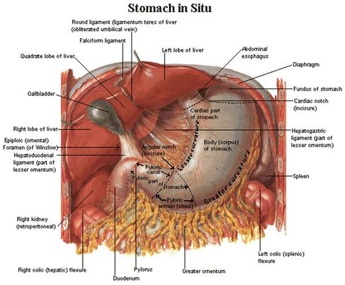
The diaphragm should also be closely inspected to rule out injury and potential contamination of the pleural cavity. It is imperative to explore the lesser sac to expose injuries on the posterior wall of the stomach. This is done most easily by gently grasping the stomach in one hand and the transverse colon in the other and lifting it up. The lesser sac is then entered by dividing the gastrocolic omentum. Completely dividing the gastrocolic omentum and taking the short gastric vessels up to the GE junction provide ultimate exposure to the stomach (Fig. 14.1). Should the suspected gastric injury not be found by visual inspection alone, the surgeon may ask the anesthesia team to fill the stomach with saline and a small amount of methylene blue dye. Gentle occlusion of the GE junction and pylorus while simultaneously applying manual pressure to the stomach can help identify the site of gastric injury.


Fig. 14.1
General anatomy and exposure of the stomach.
Gastric injuries are graded according to the American Association for the Surgery of Trauma (AAST) Organ Injury Scale (Tables 14.1, 14.2, 14.3, and 14.4). Grade I stomach injuries are contusions or hematomas or a partial-thickness laceration. A grade II stomach injury is defined as a laceration less than 2 cm at the GE junction or pylorus, less than 5 cm in the proximal one-third of the stomach, or less than 10 cm in the distal two-thirds of the stomach. A grade III stomach injury is defined as a laceration greater than 2 cm at the GE junction or pylorus, greater than 5 cm in the proximal one-third of the stomach, or greater than 10 cm in the distal two-thirds of the stomach. A grade IV stomach injury is when there is significant tissue loss or devascularization.
Table 14.1
American Association for the Surgery of Trauma (AAST)—stomach injury scale.
Stomach injury scale | ||
|---|---|---|
Grade | Description of injury | AIS-90 |
I | Contusion/hematoma | 2 |
Partial-thickness laceration | 2 | |
II | Laceration <2 cm in the GE junction or pylorus | 3 |
<5 cm in the proximal 1/3 of the stomach | 3 | |
<10 cm in the distal 2/3 of the stomach | 3 | |
III | Laceration >2 cm in the GE junction or pylorus | 3 |
>5 cm in the proximal 1/3 of the stomach | 3 | |
>10 cm in the distal 2/3 of the stomach | 3 | |
IV | Tissue loss or devascularization <2/3 of the stomach | 4 |
V | Tissue loss or devascularization >2/3 of the stomach | 4 |
Table 14.2
American Association for the Surgery of Trauma (AAST)—small bowel injury scale.
Small bowel injury scale | |||
|---|---|---|---|
Grade | Type of injury | Description of injury | AIS-90 |
I | Hematoma | Contusion or hematoma without devascularization | 2 |
Laceration | Partial thickness, no perforation | 2 | |
II | Laceration | Laceration <50 % of circumference | 3 |
III | Laceration | Laceration >50 % of circumference without transection | 3 |
IV | Laceration | Transection of the small bowel | 4 |
V | Laceration | Transection of the small bowel with segmental tissue loss | 4 |
Vascular | Devascularized segment | 4 | |
Table 14.3
American Association for the Surgery of Trauma (AAST)—colon injury scale.
Colon injury scale | |||
|---|---|---|---|
Grade | Type of injury | Description of injury | AIS-90 |
I | Hematoma | Contusion or hematoma without devascularization | 2 |
Laceration | Partial thickness, no perforation | 2 | |
II | Laceration | Laceration <50 % of circumference | 3 |
III | Laceration | Laceration ≥50 % of circumference without transection | 3 |
IV | Laceration
Stay updated, free articles. Join our Telegram channel
Full access? Get Clinical Tree
 Get Clinical Tree app for offline access

| ||


