Surgeon prefers paddle lead
Surgeon not skilled in percutaneous technique
Difficult needle access to the spine because of anatomical characteristics
Epidural fibrosis
Revision because of lead migration
Inadequate power with percutaneous leads
Positional stimulation
8.2 Technical Overview
The placement of a paddle electrode requires additional surgical skills as compared with the percutaneous placement of stimulation leads. Unlike the percutaneous cylindrical lead implantation, it does not require the ability to place a needle in the epidural space and to drive a lead. Placing a paddle lead through the open approach requires the surgical skills to safely dissect, expose the spinolaminar junction, and perform a small laminotomy, which allows for direct visualization while placing the lead (Fig. 8.1). A variety of anesthetic choices are available including general anesthesia with neuromonitoring or awake placement with local/mac or spinal anesthesia, the latter two of which still allow appropriate paresthesia mapping. While the patient is upright prior to surgery, it is important to determine the appropriate location for the future pulse generator by marking the patient’s belt line and seat line and placing the pulse generator such that it does not interfere with either. The procedure is initiated by properly positioning the patient in the prone or the semilateral position and ensuring all standard surgical precautions are taken to optimize patient safety. Once the patient is positioned, fluoroscopy is utilized to identify the appropriate interspace (e.g., T9–10).


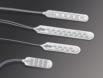
Fig. 8.1
Types of paddle leads
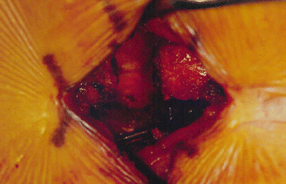
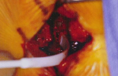
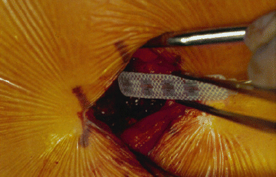
An incision is planned over the appropriate interspinous process. This area, as well as the gluteal pocket, is shaved, prepared, and draped in the usual sterile fashion (Fig. 8.2). After infiltration of local anesthetic, the thoracic incision is opened with a 10-blade scalpel. Subperiosteal dissection is performed and retractors are placed. A small laminotomy is performed (using a combination of Leksell rongeurs, Kerrison punches, and diamond drill). An adequate opening is made for the placement of the paddle lead (Fig. 8.3). Selection of the appropriate paddle lead depends on a number of factors including choice of trial system, selection of active contacts during the trial stimulation (close or wide spacing), epidural anatomy, and size of laminotomy. A paddle lead is advanced under fluoroscopic guidance such that the middle of the lead is positioned over where the patient had most adequate stimulation during the trial procedure (Fig. 8.4).


Fig. 8.2
Exposure for lead placement

Fig. 8.3
Preparation of the epidural space

Fig. 8.4
Lead placement into the epidural space
Next, the gluteal incision is opened. A pocket is made for the pulse generator and a subcutaneous pass is then made from the gluteal area to the thoracic incision. The paddle lead extension wires are brought through from top to bottom. The distal ends of the extensions are then inserted into the pulse generator and the set screws are tightened. Electronic analysis of the neurostimulator system is then performed with electrode selectability, cycling, output modulation, impedance, and compliance tested and found to be in an acceptable range.
As stated previously, some surgeons perform an awake test stimulation with a goal of obtaining paresthesias in the desired painful areas; other surgeons work under general anesthesia confirming placement using a combination of x-ray guidance and evoked potential stimulation. A typical stimulation occurs to 4–6 Hz at a pulse width of 300–350 μs with increases in amplitude until electromyographic signal changes are detected. If the goal is to achieve bilateral lower extremity stimulation, central lead placement is usually desired. In some cases, the fluoroscopic and anatomical midlines vary and the stimulation is more accurate. In cases of unilateral limb pain, the goal is to stimulate the midline and the area just off the midline to the side of pain generation.
Stay updated, free articles. Join our Telegram channel

Full access? Get Clinical Tree


