Neuropathies: diabetic, alcohol-induced, metabolic, nutritional deficiency, heavy metal, chemotherapy-induced, idiopathic, infectious (HIV, syphilis)
Spine-induced pain: disc impingement of the nerve, foraminal narrowing, central stenosis, epidural fibrosis, arachnoiditis, mechanical entrapment of the nerve, nerve trauma, iatrogenic, bone impingement on the nerve, failed back surgery syndrome
Complex regional pain syndromes types I and II, Raynaud’s syndrome, vasculitis, ischemic pain secondary to peripheral vascular disease, vasospasm
Peripheral nerve pain: nerve injury, nerve entrapment, tarsal tunnel syndrome, postsurgical scarring, neuroma, bony deformity causing nerve pain
Spinal cord stimulation (SCS) can be successful in treating this troubling problem. Conventional methods involve routine placement at the common locations in the spinal canal (T8 through T12), but in some cases, stimulation is required at the level of the nerve root or peripheral nerve. This chapter discusses possible strategies to successfully control neuropathic foot pain.
16.2 Technical Overview
Other sections of this Atlas have covered many of the pearls of this section. Needle placement, lead placement, and techniques such as anchoring and pocketing are consistent with descriptions in other sections of this text. Nerve root stimulation, paddle lead placement, and peripheral nerve placement are also covered elsewhere. The primary technical decision in treating this pain disorder is a cerebral exercise. The decision of where to target the nervous system to achieve the desired result is the most critical decision in this process (Table 16.2).
Table 16.2
Lead placement options
Approach | Location |
|---|---|
Traditional approach | T8–T12 |
Modified approach | Crossing midline at T10–T11 |
Conus approach | Crossing midline at T12–L1 |
Nerve root approach | Lead capturing the nerve root in lower lumbar spine |
Nerve root approach | Lead capturing the nerve root at foramen L4–5 or S1 |
Peripheral nerve approach | Lead placed at the peripheral nerve pain site |
Dorsal root ganglion stimulation | Dermatomal placement ipsilateral to foot pain |
16.2.1 The Method of Epidural Stimulation
Whether a percutaneous lead or a paddle lead is utilized, to achieve focally consistent and lasting stimulation of the foot, the active part of the lead should be at the T11–L1 level. This placement most likely entails stimulation of the cauda equina as it overlaps the tail end of the spinal cord, rather than the dorsal columns. The placement is often done in parallel or a staggered array (Fig. 16.1). If these lead arrays are successful, no further adaptations are needed, and in some cases, both foot pain and other dermatomal patterns are treated. Some physicians prefer to cross the midline with the leads used in a guarded array, with two cathodes in the center of the lead to drive the current deeper (Fig. 16.2). This pattern often leads to total coverage of the entire leg, including the feet. In selected patients, the paresthesia in the foot is troubling and not desired. The epidural approach is not the ideal treatment option for these patients.
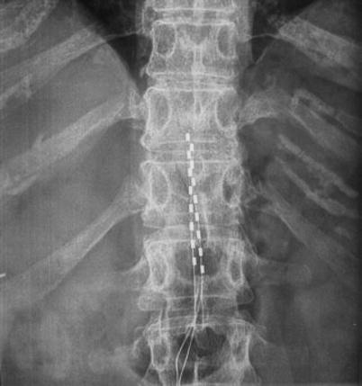
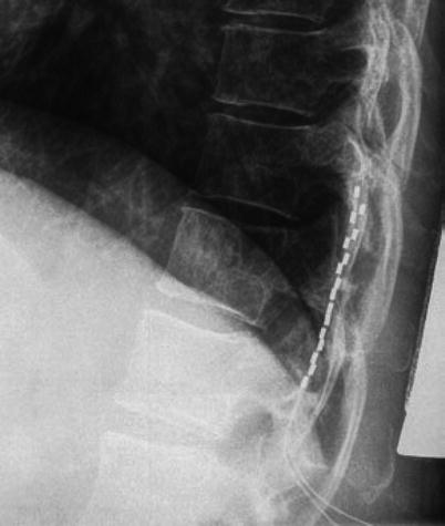
Fig. 16.1
Traditional lead placement
Fig. 16.2
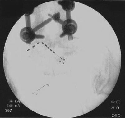
Guarded array placement. If the foot is not stimulated by the epidural approach, a nerve root approach is an option. Either a percutaneous or paddle approach can be used
16.2.2 The Nerve Root Method of Stimulation
The lead can be placed in the area of the L5 or S1 nerve root by the percutaneous approach, using the retrograde approach or the sacral hiatus route. The retrograde approach involves entering the epidural space in a caudad approach via the intralaminar space two to three levels above the target nerve. Once the epidural needle has been successfully placed with fluoroscopic guidance, the lead is driven down the middle of the epidural space until it is one level above the desired level. Using an appropriate stylet, the lead is then directed under x-ray guidance to the nerve target. The alternative method is to use a percutaneous approach to enter the sacral hiatus with an epidural Tuohy needle and then place the lead antegrade to the desired nerve foramen. The lead is placed at the foramen or adjacent to the foramen for stimulation (Figs. 16.3 and 16.4). This approach can be very helpful, but the size of conventional leads makes it difficult to stabilize this lead placement. Current research is working on developing new technology to address these issues, but currently there are no approved leads specific to the foramen or these neurological structures.