Migration risk
Physician action
Fatty tissue at anchoring site
Debride fatty tissue around the needle entry site exposing fascia and ligament for proper anchoring
Anchoring to muscle
When using an exaggerated paramedian approach, the physician should dissect medially until approaching ligament or fascia, avoiding anchoring to muscle, which may lead to migration with contraction
Lead anchor gap
The anchor should be as close to the lead entry into the ligament or fascia as possible, avoiding room for migration distal to the anchor, with newer systems allowing for a distal tracking of the catheter to “plug” the gap
Suturing with silk
Avoid silk sutures when anchoring
Dependence on the anchor
The anchor should be seen as one component of securing the system.
Total dependence on the anchor can lead to poor outcomes.
Hematoma below anchor
Hemostasis should be obtained prior to closing the wound. Bleeding can lead to catheter movement owing to hematoma compression placing pressure on the anchor.
Minimal migration changes
The catheter should be placed in an area of the spine that will not be affected by minimal migration movements. If the catheter tip is in the CSF, a good outcome may be preserved even in the presence of movement.
Once the needle and catheter are in good position, the physician must debride all fatty tissue from the area surrounding the needle, exposing the fascia and ligament. At this point the physician should place a purse-string suture around the needle. The goals of a purse-string suture are to secure the tissue that surrounds the catheter to reduce the short-term risk of CSF leak around the catheter and to reduce the risks of catheter migration in the long term by allowing the tissue to fibrose around the catheter. The purse-string is placed by using a nonabsorbable suture such as Ethibond or silk around the catheter while it is still in the needle. The suture is placed in the fascia and ligament in a purse-string orientation with at least four entries and exists is a circular pattern encompassing the needle. The suture is then tied while the needle is in place. This allows for a tight occlusion of the tissue without worry of damaging the catheter, which can result in catheter fracture or occlusion (Table 38.1).
Many anchor choices exist for physician preference in securing the catheter. Anchors are shaped in an elbow orientation, tubular form, bumpy tubular form, or butterfly shape.
Alternatives to these anchors exist with the introduction of a form-fitting bi-wing anchor on a Teflon-coated deployment device that tracks the catheter into the tissue deep to the incision.
Injection of a flowable hemostatic foam may be helpful in tissue healing and CSF leakage. The use of Gelfoam may also be helpful in reducing the risk of chronic CSF leak (Fig. 38.1a, b).
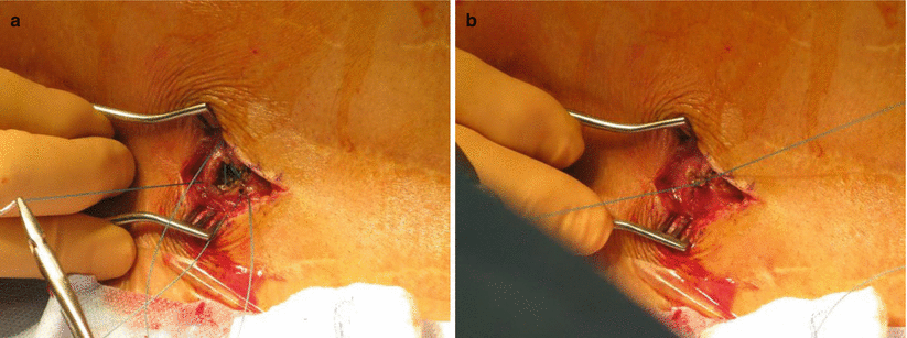
Fig. 38.1
(a) Purse-string suture. (b) “Butterfly” anchor on the catheter
Once the catheter is anchored, it is important to leave a strain-relief loop in the catheter before it is tunneled to the pocket. This will be critical in the first few weeks of tissue healing when the body movement may affect catheter security.
38.3.1 Anchor Types
Anchoring can be performed with a variety of anchors depending on manufacturer, but there are only a few basic anchor types. These include a “butterfly” anchor, which secures the catheter by wrapping around the catheter. A suture is placed through a singular suture hole to lay the catheter and anchor down against the fascia. An advantage of this type of anchor is the ability to use the same suture used for the purse-string to also secure the catheter and anchor. To accomplish this task, the suture must exit the fascia caudad to the needle entry site (Fig. 38.2).
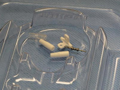
Fig. 38.2
Winged anchor with stainless steel pin and splicing catheter connectors.
The “long tubular” anchor is placed over the catheter at the proximal tip and is slid downward to the spinal entry site to abut the fascia or ligament. This type of anchor may have one, two, or three suture holes, and it is important to ensure that sutures are placed that work well in concert so that there is no strain on the catheter materials that can lead to kinking or catheter trauma.
Related posts:


Stay updated, free articles. Join our Telegram channel

Full access? Get Clinical Tree


