Agent
Effect
Ventilation pattern
Hypoventilation increases IOP, hyperventilation decreases IOP
Intravenous induction and inhaled agents
All decrease IOP except possibly ketamine; etomidate decreases IOP but myoclonus upon injection may be dangerous in setting of ruptured globe
Narcotics
Decrease IOP
Succinylcholine (Suxamethonium)
Increase IOP
Nondepolarizing neuromuscular blocking agents
Decrease IOP
Hypertonic solutions (dextran, mannitol)
Decrease IOP
Acetazolamide
Decrease IOP
Patient Evaluation
Ophthalmic surgeries are usually low-risk procedures associated with little morbidity or mortality. They are most often performed on patients at the extremes of age—the elderly and very young—both of whom merit unique considerations in the context of anesthesia. Medical history is best addressed using a systems-based approach, with a focus on the cardiovascular, pulmonary, and neurologic systems. A useful way to screen for occult cardiovascular disease is to inquire about the patient’s ability to exercise at 4 metabolic equivalents (METs) without dyspnea, chest pain, or lightheadedness. An example of an activity that uses about 4 METs is climbing one to two flights of stairs. Pulmonary evaluation should take into account recent upper respiratory infections, smoking history, and signs and symptoms suggestive of obstructive sleep apnea (e.g., snoring, obesity). The neurologic evaluation should note any preexisting deficits of the central or peripheral nervous system. The patient should also be screened for aspiration risk factors. For pediatric patients, preoperative evaluation should also include information about birth history (e.g., prematurity) and developmental history (e.g., difficulty/failure reaching developmental milestones), asthma or other respiratory problems, and recent infections. The patient’s mental status and ability to communicate adequately with the medical team is of critical importance, especially during conscious sedation cases in which the patient’s ability to remain motionless can be critical. Finally, it is important to elicit any personal or family history of complications related to anesthesia.
Laboratory testing prior to ophthalmic surgery is not usually necessary. However, hemoglobin and bleeding time may be sought for more extensive interventions such as oculofacial procedures in which blood loss is expected or in patients on medications that might affect blood clotting. Recent evidence suggests that antiplatelet and anticoagulant medications do not increase the occurrence of significant bleeding complications during cataract surgeries, even when regional blocks are performed; therefore, these medications should be continued in patients who use them chronically for cardiovascular conditions [3]. Patients with type II diabetes should be counseled to hold insulin and oral hypoglycemic agents on the day of surgery. For patients with type I diabetes, a basal infusion of insulin should be continued once nil per os (NPO), and a glucose-containing solution should be infused during surgery. A recent analysis by Operation Smile International demonstrated that the majority of children treated during its medical mission had an age–weight ratio at or below the Centers for Disease Control and Prevention’s (CDC) 50th percentile; however, the majority had near-normal hemoglobin levels. This underscores the fact that mild-to-moderate malnutrition is relatively common in developing countries. Therefore, it is best to use weight-based drug and fluid calculations [4].
Management of Anesthesia
General Principles
In developed countries, safety standards, monitoring requirements, the availability of drugs and supplies, as well as an adequate supply of well-trained anesthesia providers are such that anesthesia-related mortality is extremely low—less than 1 in 100,000 in the USA [5]. By contrast, in developing countries, deaths attributable to anesthesia in the range of 1 in 133 [6], 1 in 144 [7], and 1 in 504 [8] have been reported. To maintain a safe level of care, basic intraoperative monitoring should include means to assess oxygenation, ventilation, and circulation. Since 2007, the WHO, together with the World Federation of Societies of Anaesthesiologists (WFSA), the Association of Anaesthetists of Great Britain and Ireland (AAGBI), and others have supported the Global Oximetry Initiative to advocate the provision of pulse oximetry as a minimum monitoring standard during the provision of anesthesia [9]. Pulse oximetry offers the benefit of establishing a basic measure of adequacy of tissue perfusion and oxygenation as well as providing a continuous display of heart rate. When general anesthesia is administered, continuous capnography is the gold standard for ensuring correct placement of the airway and adequacy of ventilation. An esophageal or precordial stethoscope provides an alternative means of assessing adequacy of ventilation when capnography is not available. The WFSA has published recommendations on international standards for the safe practice of anesthesia based on available levels of infrastructure (basic, intermediate, optimal) [10]. This includes suggested minimum drug requirements (Table 15.2). The WHO also has a Model List of Essential Medicines [11].
Table 15.2
Recommended supplies and anesthesia drugs based on level of health care facility
Rural hospital or health center (sparse operating room or minor procedure room only) | District or provincial hospital (100–300 beds) | Referral hospital with intensive care facilities (300–1,000 beds) |
|---|---|---|
Supplies | ||
Equipment: Capital | ||
Complete anesthesia, resuscitation and airway management systems including: | Same as Level 2 with these additions per operating room and ICU bed: | |
Adult and pediatric self-inflating breathing bags with masks | Reliable oxygen source | ECG monitor |
Foot-powered suction | Vaporizer | Anesthesia ventilator, reliable electric power source with manual override |
Stethoscope, sphygmomanometer, thermometer | Hoses and valves | Infusion pumps |
Pulse oximeter | Bellows or bags to inflate lungs | Pressure bag with IV infusion |
Oxygen concentrator or tank oxygen and draw-over vaporizer with hosesa | Face masks (size 00–5) | Electric or pneumatic suction |
Laryngoscopes, bougies | Pediatric anesthesia system | Oxygen analyzer |
Oxygen supply failure alarm, oxygen analyzer | Thermometer or temperature probe | |
Adult and pediatric resuscitator sets | Electric warming blanket | |
Pulse oximeter, spare probes, adult and pediatric | Electric overhead heater | |
Capnograph | Infant incubator | |
Defibrillator (one per OR suite/ICU) | Laryngeal mask airways (sizes 2–4) | |
ECG monitor | Intubating bougies, adult and child | |
Larygnoscope, Macintosh blades (1–4) | Anesthetic agent (gas and vapour analyzer) | |
Oxygen concentrator[s] cylinder | ||
Foot or electric suction | ||
IV pressure infusor bag | ||
Adult and pediatric resuscitator sets | ||
Magill forceps and/or bougie | ||
Spinal needles (25G) | ||
Nerve stimulator | ||
Automatic noninvasive blood pressure monitor | ||
Equipment: Disposable | ||
Examination gloves | ECG electrodes | Same as Level 2 plus: |
IV infusion/drug injection equipment | IV equipment and fluids (normal saline, Ringer’s lactate, dextrose 5 %) | Ventilator circuits |
Suction catheter size 16F | Pediatric giving sets | Yankauer circuits and suckers |
Airway support equipment, including airways and tracheal tubes | Suction catheter size 16F | IV infusion tubing |
Oral and nasal airways | Sterile gloves sizes 6–8 | Disposable suction machines |
Nasogastric tubes 10–16F | Disposables for capnography, oxygen analyzer | |
Oral airway size 000–4 | Sampling lines | |
Tracheal tube size 3–8.5 mm | Water traps | |
Spinal needle size 22G and 25G | Connectors | |
Battery size C | Filters—Fuel cells | |
Drugs | ||
Ketamine 50 mg/mL | Same as Level 1 but also: | Same as Level 2 but also: |
Lidocaine 1 or 2 % | Thiopental 500 mg/1 g powder or Propofol | Propofol |
Diazepam 5 mg/mL or Midazolam 1 mg/mL | Suxamethonium bromide 500 mg powder | Nitrous oxide |
Morphine 10 mg/mL | Pancuronium | Various modern neuromuscular blocking agents |
Pethidine (Meperidine) 50 mg/mL | Neostigmine 2.5 mg injection | Various modern inhalation agents |
Epinephrine 1 mg | Ether, halothane, other inhalation agents | Various inotropic agents |
Atropine 0.6 mg/mL | Lidocaine 5 %, heavy spinal | Various antiarrhythmic agents |
Bupivacaine 0.5 %, heavy or plain | Nitroglycerine for infusion | |
Hydralazine 20 mg injection | Calcium chloride 10 % 10 for injection | |
Furosemide 20 mg injection | Potassium chloride 20 % 10 mL injection for infusion | |
Dextrose 50 % 20 mL injection | ||
Aminophylline 250 mg injection | ||
Ephedrine 30/50 mg ampule | ||
Hydrocortisone | ||
±Nitrous oxide | ||
For ophthalmic procedures, maximizing the use of local and regional anesthesia offers the benefit of minimizing equipment and monitoring needs. It is therefore an ideal approach for the majority of cases. It should be recognized that although it is possible for a visiting medical mission group to transport disposable supplies and drugs, the team will often be dependent on the host nation for daily sterilization of equipment. Therefore, the quality of this sterilization equipment may be a limiting factor in terms of case turnover. It is advisable to provision supplies for at least a full day of surgical procedures without needing to re-sterilize equipment. Carrying disposable back-up instruments can also provide a safety net in the event reusable instruments become unavailable.
In some countries, traditional NPO guidelines are circumvented, especially for those patients who are only expected to receive regional ophthalmic blocks or “minimal” sedation [12]; however, this practice is controversial, and following the ASA Practice Guidelines on Preoperative Fasting is the safest approach (Table 15.3) [13].
Table 15.3
Summary of fasting guidelines to prevent pulmonary aspiration
Ingested substance | Minimum fasting period (h) |
|---|---|
Clear liquids (water, carbonated beverages, tea, black coffee) | 2 |
Breast milk | 4 |
Infant formula | 6 |
Nonhuman milk | 6 |
Light meal (toast, clear liquids) | 6 |
Heavy meal (fatty foods) | 8 |
As with any anesthetic, it is useful to utilize a pre-anesthetic checklist to ensure that basic preoperative patient information and the necessary anesthesia equipment is available prior to the start of each case (Table 15.4 and Fig. 15.1).
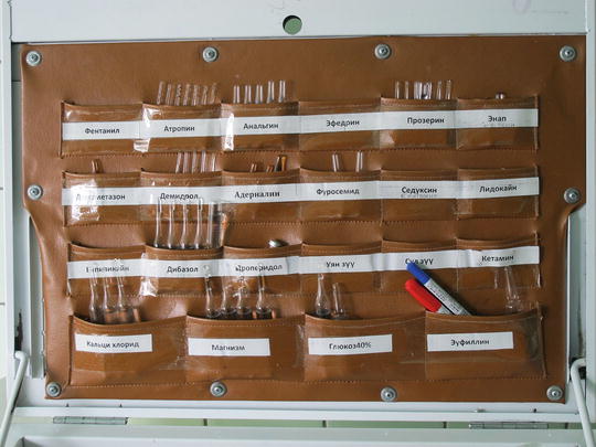
Table 15.4
Pre-anesthetic checklist
Patient’s Name: Date of Birth: Weight: | Procedure: | ||
|---|---|---|---|
ASA Physical Status | 1 2 3 4 5 E | Anesthetic resources | |
Mallampati Class | I II III IV | Airway – Masks – LMAs, Tubes – Working laryngoscopes – Bougies | ⃞ ⃞ ⃞ ⃞ |
Aspiration Risk? | Yes No | Breathing – Circuit, leak-tested – Soda lime, no color change | ⃞ ⃞ |
Allergies? | Yes No | Suction | ⃞ |
Important Medications (e.g., insulin, seizure drugs) | Drugs and Devices – Oxygen cylinder, full and off – Vaporizers – Monitors, alarms on – Based drugs labeled – Fluids available – Thermometer, temperature probe | ⃞ ⃞ ⃞ ⃞ ⃞ ⃞ | |
Relevant Medical Problems (e.g., diabetes, asthma) | Emergency – Epinephrine – Self-inflating bag – Tilting table – Succinylcholine | ⃞ ⃞ ⃞ ⃞ | |
History of anesthesia-related problems | |||
Fig. 15.1
When relying on a local supply of medications, it is imperative to foresee a mechanism to identify each drug appropriately
Regional Blocks
Topical local anesthesia and regional orbital blocks may be employed for most intraocular and periorbital procedures. The increased use of phacoemulsification for cataract extraction has led to a greater use of topical anesthesia for cataract surgery, while most vitreoretinal procedures require a denser anesthesia provided by regional blocks [14]. Among the regional techniques available are the retrobulbar, peribulbar, and sub-Tenon’s block. Relative contraindications to regional ophthalmic block include blindness in the contralateral eye, significant myopia, elevated IOP, and trauma or perforation of the globe [15]. Whether these blocks are performed by an anesthesiologist or ophthalmologist varies by institution and experience of the provider. Regardless, it is the responsibility of the anesthesiologist to monitor the patient’s vital signs during the performance of any regional anesthetic.
Retrobulbar and Peribulbar Blocks
Traditionally, the four rectus muscles of the eyes were thought to form a distinct cone around the globe whose apex was at the optic foramen. Retrobulbar blocks are performed such that local anesthetic is injected directly into the intraconal space, allowing for profound and rapid anesthesia and akinesia (globe immobility) of the rectus muscles with a low volume of anesthetic. Subsequently, it has been recognized that the connective tissue around the rectus muscles is incomplete. Therefore, local anesthetic injected outside the orbital cone may diffuse into the intraconal space and produce results similar to those achieved from a retrobulbar block with relatively less risk. This is the principle behind the peribulbar block, which is theoretically safer owing to the position of the needle on injection more removed from the optic nerve and other critical brainstem structures.
Related posts:
 The Evolution of Surgical Humanitarian Missions
Trauma, War, and Managing Vascular and Orthopedic Injuries
Surgical Mission Trips as a Component of Medical Education and Residency Training
The Evolution of Surgical Humanitarian Missions
Trauma, War, and Managing Vascular and Orthopedic Injuries
Surgical Mission Trips as a Component of Medical Education and Residency Training
 Legal and Ethical Issues in Global Health: A Trip Through the Vagaries of Truth and Culture
 Medical Missions: A Short History from There to Here
Stay updated, free articles. Join our Telegram channel

Full access? Get Clinical Tree


