Authors, Year, Type of study
Studies, Number of participants
Type of patients
Monitoring devices
Conclusions
Poeze et al. [9], Meta-analysis
30 RCTs, 5,733 pts
High-risk surgery, trauma, acute medical sepsis
PAC, esophageal Doppler ultrasonography
Hemodynamic optimization improves mortality in perioperative and trauma setting. Overall trial quality is moderate
Brienza et al. [10], Meta-analysis
20 RCTs, 4,220 pts
High-risk surgical patients
PAC, lithium dilution, analysis of arterial waveform, esophageal Doppler ultrasonography
Hemodynamic optimization prevents post-operative renal dysfunction in high-risk patients Mortality was reduced in perioperative setting
Gurgel et al. [11], Meta-analysis
32 RCTs, 5,056 pts
High-risk surgical patients
PAC, esophageal Doppler ultrasonography, analysis of arterial waveform, lithium indicator dilution, thoracic bioimpedance
Maintenance of tissue perfusion with a specific protocol improves outcome and reduces mortality in high-risk surgical patients when PAC was used. CI, DO2, and VO2 are the most useful target for hemodynamic management
Hamilton et al. [12], Meta-analysis
29 RCTs, 4,805 pts
Moderate-high-risk surgical patients
PAC, esophageal Doppler ultrasonography, analysis of arterial waveform, lithium indicator dilution
A preemptive-targeted approach to the hemodynamic in perioperative period may reduce morbidity and mortality after surgery in high-risk surgical patients. Very few studies were performed in high-quality design
Finally, all meta-analysis considered show a “grey area” given by the methodological quality of the individual studies examined. In fact, studies turned out to be of moderate quality, limiting the significance of their findings to high-risk surgical patients. Single-centered, non-blind studies and with inadequate statistical power for the sample size do not show significant effects on moderate-risk patients and for less invasive monitoring systems.
6.3 Physiopathology
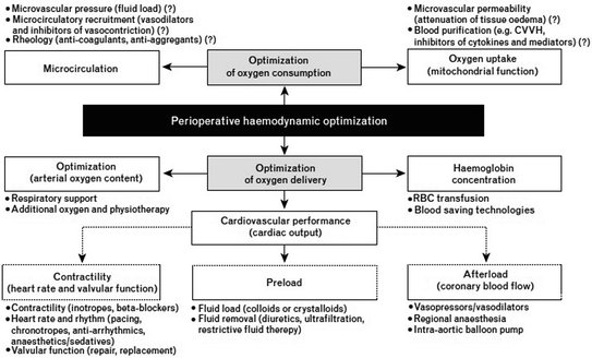
All studies examined in the Consensus Conference by Landoni and others share the common physiopathological background of maintaining adequate tissue oxygenation [3]. It is a well-known fact that surgery is associated with a systemic inflammatory response with increased oxygen consumption (VO2). In high-risk surgical patients (expected mortality >5 %), whose characteristics are summarized in Table 6.2, the cardiovascular system cannot counterbalance this increased oxygen consumption with adequate delivery. The oxygen debt thus created plays a role in tissue hypoxia, which results in post-operative complications and, in some cases, death [13]. Specifically, in the intestine, reduced oxygen perfusion causes damage to the endothelial barrier with the release of endotoxins into blood circulation; these endotoxins activate and stimulate the multi-organic inflammatory response [4]. Hence, it is explained how maintenance of renal function and prevention of tissue hypoperfusion are strictly related to perioperative survival. Tissue hypoxia, at an early stage, is “hidden,” as it cannot be diagnosed through common clinical parameters such as mean arterial pressure, heart rate (HR), or central venous pressure. Even less useful are early clinical signs such as diuresis, state of consciousness, and peripheral perfusion. Prevention of tissue hypoxia depends on several factors and involves a balance between oxygen delivery and consumption (Fig. 6.1). While oxygen consumption can be only minimally modified, its delivery is the key element in hemodynamic optimization. It is given by the product of the following parameters:

Table 6.2
Clinical criteria for high-risk surgical patients requiring perioperative hemodynamic optimization (from Kirov [16])
Patient-related criteria | Surgery-related criteria |
|---|---|
Severe cardiac or respiratory illness resulting in severe functional limitation | Extensive non-cardiac surgery (e.g., carcinoma involving bowel anastomosis, pneumonectomy, complex traumatological and orthopedic procedures) |
Aged over 70 years with moderate functional limitation of one or more organ systems | Major/combined cardiovascular surgery (e.g., aortic aneurysm, combined valve repair, coronary surgery, and carotid endarterectomy) |
Acute massive blood loss (>2.5 l) | Surgery prolonged >2 h (e.g., neurosurgical interventions, combined gastrointestinal surgery) |
Severe sepsis | Emergency surgery |
Shock or severe hypovolemia of any origin | |
Respiratory failure (paO2 <60 mmHg or SpO2 <90 % in spontaneously breathing patients receiving oxygen or paO2/FiO2 <300 in mechanically ventilated patients or ventilation >48 h) | |
Acute gastrointestinal failure (e.g., intraabdominal compartment syndrome, pancreatitis, perforated viscus, gastrointestinal bleeding) | |
Acute renal failure (urea >20 mmol/l, creatinine >260 mcmol/l) |

Fig. 6.1
Concept of perioperative hemodynamic optimization (from Kirov et al. [16]) (CVVH continuous veno–venous hemofiltration)
DO2 (mL/min) = cardiac output (CO) (L/min) × arterial oxygen content (CaO2)
While you need to preserve arterial oxygen content by maintaining adequate hemoglobin and blood oxygen level, a key element of hemodynamic optimization is given by the CO or CI, as it can be rapidly monitored and adapted to the patient’s bedside. Different systems of hemodynamic monitoring (Table 6.3) allow, with different methods depending on their technology and invasiveness, to estimate the stroke volume (SV) and, from this, to obtain the cardiac output value (CO = SV × HR). Besides this perfusion index, current devices allow for a more precise and focused hemodynamic management through the measurement of other parameters:
Methodology | Cardiac output | Static preload data | Dynamic preload data | Oximetry data SvO2/ScvO2 | Limits | Notes | |
|---|---|---|---|---|---|---|---|
Expanded invasive monitoring | Pulmonary artery catheterization | Continuous, delayed response time of several minutes | CVP PCWP | – | SvO2 | Operator dependence Patients conditions (mitral or tricuspid valve insufficiency, shunt) | Gold standard |
Less invasive | Calibrated pulse pressure analysis | Continuous, rapid response time (s) | CVP, GEDV, EVLW | SVV, PPV | ScvO2 | Arrhythmias, IABP. Peripheric arteriopathy frequent recalibration during severe hemodynamic instability | Static preload data |
Minimally invasive
Stay updated, free articles. Join our Telegram channel
Full access? Get Clinical Tree
 Get Clinical Tree app for offline access

|

