Fig. 25.1
Anatomy of fingertip in sagittal section. Lateral view of finger depiction dorsal aspect to volar aspect of nail, nail bed, distal phalanx, and volar tip from distal proximal phalanx to distal tip
The fingertip has a radial and ulnar digital nerve which has three distinct branches distal to the distal interphalangeal joint. The radial and ulnar digital arteries provide the dominant flow and are situated on the volar aspect of the finger and run with the digital nerves. The veins are predominantly dorsal and their location prevents venous congestion with pinch and grasp. The extensor tendon attaches to the proximal distal phalanx on the dorsal aspect. The flexor digitorum profundus tendon attaches to the proximal volar aspect of the distal phalanx. Injuries to any of these structures can warrant specialized care but most fingertip injuries do not require nerve or tendon care. An amputation proximal to the germinal matrix and nail fold requires neurectomies or tendon release as the flexor and extensor tendons should not be sewn together to prevent the quadregia effect and impair digital function.
Patient Evaluation
Evaluation of a patient with a fingertip injury begins with a thorough history which should include the mechanism of injury. A sharp and clean injury may be more amenable to an anatomic reconstruction than a dirty, crush, or avulsion injury. The presence or absence of the injured part determines whether replantation or use as a composite graft is possible. Patient factors to consider include hand dominance, age, occupation, recreational pursuits, history of previous injuries or hand problems. Medical comorbidities such as cardiac or vascular disease, diabetes, smoking, and alcohol consumption should also be assessed. A complete examination of the hand includes an assessment of the skin, vascularity, neurologic function and flexor and extensor tendon function. The injury is inspected with specific attention to the characteristics of the wound. Tetanus status should be assessed and cultures obtained in grossly contaminated injuries such as a barnyard injury. Intravenous antibiotics are indicated for patients with an associated fracture.
Inspection of the hand starts with color. Ischemia will cause a loss of the normal pink vascular appearance of the nail and volar tip. The nails should be smooth and the nail bed pink as well. Compression should cause a blanching that should clear in 1–3 s with a return to a pink color. Venous congestion can cause the tip and nail to turn blue and even darker in color. Infarcted or necrotic tissue is black. Hemorrhage in the nail bed can be a harbinger of fracture of the distal phalanx and a nail bed injury. The finger has a turgor to it that should be symmetric amongst all digits. The volar tip has a set of ridges that comprise our fingerprint. Each finger is unique with respect to the pattern of ridges (fingerprint) and care should be taken to minimize incisions and painful scars over the volar fingertip and pad.
After gross inspection of the soft tissue injury, radiographs of the hand and finger should be obtained to assess the change in the boney architecture. A clean amputation with bone involvement will have fewer possible issues to consider than a comminuted fracture involving the proximal distal phalangeal articular surface and possibly the middle phalanx distal articular surface.
Injury Patterns and Classification
Mechanisms of injury vary and range from clean sharp trauma to dirty blunt and crush injury. The mechanism of injury can be a guide to treatment. A sharp injury with an available soft tissue piece can be defatted and placed as a skin graft or sewn back on as a composite graft. If the soft tissue piece is gone and there is not any exposed bone, closure or hypothenar split thickness skin graft could be possible. An injury to bone and nail bed may require shortening and closure or a reconstructive local flap such as a V–Y flap or lateral Kutler flap. More complex techniques such as the vascular island flap, thenar flap, cross finger flap, or groin flap closure would likely require transfer to a hand surgical specialist.
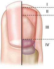
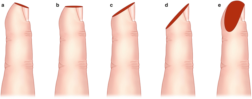
Fingertip injuries can be divided into crushing injuries or clean amputations and can be classified based on the level of the amputation, the obliquity of the wound, and whether there is any exposed bone (Fig. 25.2). The Allen Classification is commonly utilized to describe fingertip injuries and can be used as a guide for treatment. Type I injuries are those in which only the pulp of the finger is involved without any exposed bone. Type II injuries are those in which there is both pulp and nail loss as well as exposed bone. Type III injuries are those in which there is partial loss of the distal phalanx as well as both pulp and nail loss. Type IV injuries are those in which the lunula of the nail is involved along with the pulp, nail bed, and distal phalanx. The obliquity of the amputation wound can be described as dorsal oblique, transverse, volar oblique, or lateral oblique (Fig. 25.3).


Fig. 25.2
Allen classification of fingertip Injuries

Fig. 25.3
Obliquity of the amputation
In Allen Type I injuries of the fingertip where there is loss of skin or pulp tissue without exposed bone, the wound may be allowed to heal via secondary intention or by performing a skin graft. Controversy exists as to which method is better, but most agree that wounds larger than 1 square centimeter are better treated with full-thickness skin grafting. In Type II and III injuries, with bone exposure, the wound may be closed primarily or a local flap may be utilized. Type IV injuries which are proximal to the lunula, can be treated by revision amputation, flap coverage, or microsurgical replantation. When bone is exposed, as in Type II–IV, the question becomes whether length should be preserved, necessitating coverage, or if sacrifice of length is appropriate.
Preoperative Preparation
The patient should be carefully positioned with a hand table or appropriate substitute table to allow for placing the hand on a flat surface for exposure. The hand table can be a specialized table that allows for preparation and draping of a sterile field. A mayo stand or arm board may suffice if a hand surgery table is not available. Other injuries may require primary care as part of a primary and secondary survey. A digital or arm tourniquet should be available and used as necessary to minimize bleeding and optimize visualization. A good light source should be positioned. Adequate magnification may be helpful such as surgical loupes. The surgeon should consider a good irrigation and debridement of the wounds with a digital or wrist block anesthesia. Antibiotics should be administered and considered in the postsurgical period for 48–72 h. Open fractures and grossly contaminated wounds may require a longer period of up to 7–10 days. A sterile field should be set with adequate head, eye and mask coverage for the surgeon. Sterile hand washing, sterile gloves and perhaps a gown must be employed. Adequate instruments, suture, Adaptic, Xeroform, and dressings should be immediately available. Careful planning is essential if an assistant is not available to obtain materials as the reconstructive procedure moves forward.
After the evaluation of the patient and wound is completed, a treatment plan is formulated. If more than one option is available, the advantages and disadvantages of each should be discussed with patient, and the simplest method that accomplishes the desired result should be pursued. Most fingertip injuries can be managed in the emergency department, but complex regional flaps as well as more invasive procedures are more appropriately treated in the operating room.
Facility and equipment availability requires consideration. Appropriate instruments including a freer elevator, rongeur, proper knife blades, and suture are necessary to technically perform the reconstructive procedure. A supply of dressings and splints are essential for postsurgical care. Access to follow-up care will optimize patient satisfaction and outcomes.
Debridement and Closure
Operative Strategy
Primary closure of a distal tip amputation of pulp may be an option where the location and amount of skin loss allow for closure without excessive tension. There must be adequate tissue for closure or shortening of the distal bone is necessary.
Operative Technique
The technique will differ depending on the nature of the injury and the complexity of the wound. In most cases, the digit may be anesthetized using a digital block. Four nerve branches, two dorsal and two volar, supply each digit. The dorsal nerves should be blocked only if the intervention will extend proximally from the distal interphalangeal joint. A 25-gauge with 10 cm3 of 1 % lidocaine, or 0.5 % marcaine if longer duration is needed, can be inserted at the level of the distal palmar crease and 2.5 cm3 of anesthetic can be injected on both sides of the metacarpal neck. A circumferential ring block at the base of the digit is not recommended because the subsequent pressure may result in ischemia to the digit.
The wounds should be effectively irrigated and debrided of devitalized tissue. A hematoma area 50 % of the size of the nail indicates a significant nail bed injury. Drainage with a sterile 18-gauge needle can alleviate pain. If a nail bed injury is present, the nail plate can be removed, and the sterile matrix should be re-approximated with 6-0 chromic gut or similar absorbable suture. After nail bed repair is completed, the nail plate should be placed into its native position to maintain the nail fold and protect the repair. The skin can be closed with a 4-0 or 5-0 nylon or similar sized non-absorbable monofilament suture. Absorbable sutures can be used in children or in places when removal may be a challenge. A sterile non-adherent dressing is applied. The finger should be immobilized.
Stay updated, free articles. Join our Telegram channel

Full access? Get Clinical Tree


