Chapter 12 Regional complications in joint hypermobility syndrome
12.1 The shoulder joint
Laxity
Inappropriate laxity, energy expenditure and the rotator cuff
The quality of proprioception is reduced in shoulders with hyperlaxity, instability, and previous capsular trauma. Improvement in joint position and motion sense has been shown in trained shoulders and after anterior capsular reconstruction in those with traumatic structural instability (Myers et al 2006). It is not clear whether this is also the case for patients with atraumatic structural instability.
Common disease conditions
Glenohumeral instability
Classification
The relative importance of each factor to the syndrome can change over time. The pathologies causing instability comprise structural (RC, SAC, CLPM) and non-structural (NS) elements (Lewis et al 2004). The structural elements may be congenitally abnormal, comprise abnormal collagen, acquire microtraumatic lesions over time (atraumatic structural) or be damaged by extrinsic force (traumatic structural). The non-structural elements can be congenitally abnormal or acquired over time as perturbations of neuromuscular control, particularly at periods of skeletal growth.
The concept of instability (which may change over time) being caused by a combination of structural (traumatic and atraumatic) and neurological system disturbances, has led to the classification of instability as a continuum of pathologies, which can be graphically displayed as a triangle (Lewis et al 2004). The polar pathologies are labelled type I (traumatic instability), type II (atraumatic instability) and type III (neurological dysfunctional or muscle-patterning). Polar groups I and II and the axis I–II, representing the spectrum between the two poles, correspond to the TUBS-AMBRI classification (Thomas & Matsen 1989). This allows for a spectrum of structural shoulder pathology but does not admit those shoulders in which there is no structural (traumatic or atraumatic) cause for the instability. Personal observation has further subdivided polar group III into peripheral, central, protective and combination subtypes (A Jaggi, personal communication). The interpolar spectrum describes dual pathologies in which traumatic, atraumatic and muscle-patterning factors play a variable role in the emergence of instability. All groups can be represented in patients with JHS, but more commonly present in groups II and III.
Presentation
JHS patients may present with pain rather than overtly abnormal displacements. Anterior atraumatic instability is less common than posterior types (Malone et al 2006a). Posterior structural anomalies (including posterior glenoid dysplasia, excessive glenoid retroversion, glenoid hypoplasia and medialized posterior capsular attachment) create the environment in which obligatory positional posterior displacement may occur during elevation of the arm in the sagittal plane. If abnormal muscle activation appears clinically obvious, either at the onset of motion, or during the motion, then the diagnosis has a muscle patterning component (type III). If there is no clinically or electrophysiologically proven aberrant muscle activation then the condition is labelled type II (positional subtype).
Muscle-patterning instability (MPI) comprises aberrant activation of large muscles and simultaneous suppression of the rotator cuff. Thus far, dynamic electromyography has characterized latissimus dorsi, pectoralis major and anterior deltoid among the large muscles, and only infraspinatus of the rotator cuff. It is as yet not clear whether suppression of infraspinatus reflects whole rotator cuff suppression or specific suppression of the infraspinatus. MPI can be clinically obvious in polar group III, but may be occult, requiring dynamic electromyography (DEMG) for confirmation in type II/III and III/II instabilities (Malone et al 2006b).
Clinical assessment
Abnormal muscle patterning is the result of aberration in one or more of three sub-systems, which contribute to neuromuscular control. At a peripheral level, capsular laxity and abnormal collagen composition may affect proprioception and lead to injury and resultant instability (Blasier et al 1994, Myers & Lephart 2002). Furthermore, a proprioceptively deficient joint can disrupt movement at other joints along the kinetic chain by altering the motor programme, thereby affecting control at a central level (Myers & Lephart 2002). In addition, impaired central nervous motor programming due to stress, constrained movements, postures and/or chronic fatigue will influence muscle tone, creating the risk of instability. Assessment of all three systems is therefore necessary to aid appropriate management. (Chapter 3, Fig. 3.2 and Chapter 8, Fig. 8.1 & 8.2)
Subjective assessment
The mechanism of the event that causes the initial symptoms is key to classification. JHS patients will commonly describe atraumatic events. These can include an over-stretching manoeuvre or repetitive use, which causes initial displacement but with spontaneous relocation or relocation with minimal assistance. Impaired proprioception leads to a propensity to injury and the most minor incidents can cause easy displacement of the glenohumeral joint (GHJ) with minor or no resultant structural damage. Subsequently, the patient notices recurrent episodes of dislocation on simple manoeuvres often associated with daily activities. Some patients with a predominant muscle-patterning problem often describe an ability to voluntarily displace the shoulder, a ‘party trick’. An initial childhood voluntary trick causing no problems, may develop after minor injury, into an involuntary problem creating a so-called habitual problem. On occasion, the shoulder can remain persistently displaced as a result of ongoing abnormal muscle activity. Patients may recall that emergency room staff had difficulty relocating the shoulder or keeping the joint located after cessation of anaesthesia. During muscular relaxation under anaesthesia, the shoulder reduces spontaneously, but on recovery of conscious function the shoulder re-dislocates readily, but, importantly, not voluntarily. How much of this ongoing abnormal muscle activation is a result of pain, fear and altered central inhibitory control (Barrett et al 2000) is difficult to ascertain, but points to the likelihood of a non-structural cause to the instability.
In adolescents, accelerated growth may be associated with exacerbation of symptoms (Chapter 10). The peak incidences of new-onset muscle-patterning instability occur at about 10, 15 and 20+ years, suggesting a coincidence with pubertal hormonal effects, especially within the female cohort (Malone et al 2006a). Leisure and sport activities should also be considered. The repetitive nature of an activity such as swimming may expose the predisposed joint to overuse, leading to peri-articular muscle fatigue. Certain activities, such as poorly supervised weight-training may cause asymmetric overuse in agonist muscle groups, thus exacerbating postural and motor imbalances. A common pattern involves a dominance of powerful internal rotators (pectoralis major and latissimus dorsi) over weaker, or suppressed, external rotators (infraspinatus) and aberrant trunk stability.
Psychological stress has been implicated in the production of symptoms. However, experience at our unit strongly suggests that the observed stress and anxiety is secondary to the persistent challenge of painful instability. Fear of using the upper limb or modification of activity compounds the problem, altering central motor control. Careful and sensitive history-taking allows the therapist to extract this information and avoid the implication that patients may be displacing their shoulder for a secondary gain, which has sometimes been implied by health professionals who do not appreciate that muscle patterning can contribute to shoulder instability. Involvement of clinical psychologists can be useful to help the patient acknowledge and manage their fear of, as well as the fact of, persistent and recurrent instability and prepare the path for active management of JHS (Chapter 8).
Objective assessment
A thorough clinical examination must not only assess the shoulder, but also include posture, global laxity, scapula dysrhythmia, general balance and proprioception (Chapter 6.4). The shoulder does not function in isolation, but provides the integral link in producing and transferring energy from the trunk to the arm (Kibler et al 2001). Lower limb and trunk stability has a resultant effect on the scapula and GHJ. Integrating multiple body segments (proximal to distal) rehabilitates the entire neuromuscular system, so optimizing shoulder function (McMullen & Uhl 2000).
Posture
An assessment of standing posture should be performed from all directions. The hypermobile patient typically adopts the sway-back posture as described by Kendall et al (1993), a tendency to hyperextend the hips and knees and cause forward displacement of the pelvis. This tendency to hang at the extreme of their hypermobile range results in poor core stability and may increase the use of more superficial torque muscles such as latissimus dorsi, having a resultant effect at the shoulder girdle (Gibson 2004).
A secondary thoracic kyphosis may develop to compensate for the sway posture. As a result the scapula adopts a protracted, laterally tilted position. The glenoid cavities face downwards and forwards, such that the humeral head ‘hangs’ from the glenoid, further challenging the joint reaction compression force. If the medial border or inferior pole of the scapula also wings, the combination of inhibition of the scapula stabilizers (serratus anterior and the lower fibres of trapezius) with increased activity in the pectoral and latissimus muscles (Kibler 2000a, Mottram 1997) should be assessed.
Quality of movement
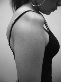
Aberrant scapular mechanics are fundamental to the aetiology of instability and must be assessed both at rest and dynamically. The scapula is observed as the patient elevates the arm in both flexion and abduction planes. It is also useful to assess scapular stability in weight bearing by placing the patient in four-point kneeling. This may highlight scapula winging as well as poor trunk stability (Magarey & Jones 2003) (Figure 12.1.1). Movement faults are not always present on one manoeuvre and if symptoms tend to be present on overuse, it is helpful to ask the patient to repeat movements with varied speed to pick up abnormality.
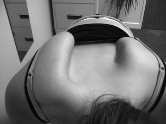
In those patients who can demonstrate a party trick it is useful to observe the direction of displacement and any associated abnormal muscle activation. It is also important to observe the resting position of the GHJ prior to movement, to be sure it is not already displaced under the effect of abnormal resting tone in surrounding muscles. A persistently inferiorly displaced GHJ can prove difficult to manage, because this indicates gross inhibition of rotator cuff and deltoid tone which is likely to be centrally driven (Figure 12.1.2).
Patients with MPI usually present with posterior instability (Takwale et al 2000, Kuroda et al 2001, Malone et al 2006a). The dominant pattern of elevation of the arm is in internal rotation (associated with overactivity in latissimus dorsi and/or pectoralis major), with resultant or associated inhibition of infraspinatus and posterior deltoid. As a result, posterior displacement of the GHJ occurs in ascent with an associated ‘reverse-scapular’ action, as the scapula is prevented from protracting and upwardly rotating in the normal way (Takwale et al 2000). As the arm descends, there may be an audible click or clunk as the GHJ relocates.
A useful clinical test to confirm whether posterior instability is a result of such imbalance is the external rotation facilitation test. The practitioner facilitates the action of infraspinatus by encouraging active external rotation against resistance as the limb is elevated. If the GHJ remains stable (located) then treatment should be focused towards restoring normal movement patterns (Takwale et al 2000) through biofeedback programmes, which facilitate infraspinatus and suppress pectoralis and latissimus activity.
Muscle activation
Conservative management of shoulder instability has concentrated on strengthening exercises to the rotator cuff (Aronen & Regan 1984, Burkhead & Rockwood 1992). However, it is now recognized that abnormal muscle activation in other muscle groups can contribute to instability. Experience at our specialist unit through the use of DEMG has indicated that abnormal activation patterns in latissimus dorsi, pectoralis major, anterior deltoid and infraspinatus contribute to shoulder instability (Malone et al 2006b). In the absence of electromyography equipment, the therapist can assess abnormal tone through palpation and observation as discussed above. Asking the patient to perform simple distal movements with the hand or elbow, and palpating for any increased activation of the pectoral or latissimus dorsi muscles, may indicate abnormal recruitment. Strengthening programmes may not always be fruitful in the presence of abnormal tone in other muscles and sometimes management may need to focus on inhibiting muscle activation to gain stability.
Standard instability tests for the shoulder may be useful in cases where observation alone does not indicate the direction of instability, but it must be recalled that asymptomatic translation of the glenohumeral joint simply indicates laxity of the joint. Instability can, by definition, only be present if the patient has recognizable symptoms during the provocative manoeuvre. JHS patients can present with clumsiness, poor co-ordination and gross motor maldevelopment (Adib et al 2005), so additional neurological tests to assess cerebellar, basal ganglia and dorsal (spinal cord) column functions are useful. General balance and stability can be tested by the single-leg balance test, both with eyes open and closed (Chapter 6.4). Poor control will highlight lower limb and trunk imbalances, which may be contributing to the instability (Kibler et al 2001, Gibson et al 2004).
Management
Correcting abnormal muscle patterns and appropriate strengthening are the key principles in management. Re-establishing neuromuscular control must integrate peripheral somatosensory, visual and vestibular afferent input and improve motor control through spinal reflex, brain stem and cognitive programming (Lephart & Henry 2000, Griffin and Letha 2003).
Early and intermediate stages
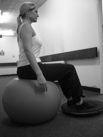
Postural awareness and correction is the foundation of early intervention. Improving core strength may directly influence abnormal muscle tone at the shoulder girdle. This can be achieved by simple postural exercises such as back flattening against a wall, challenging balance using a Swiss ball (Fig. 12.1.3) or balance board and single-leg stance to increase postural tone. Research has shown that preparatory trunk muscle activation accompanies limb motion (Zattara & Bouisset 1988, Hodges et al 2001) and supports the concept of working on posture prior to upper limb movement.
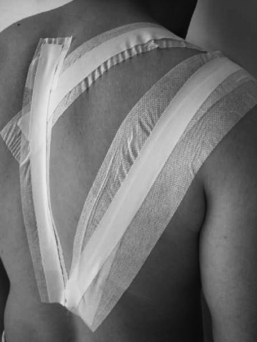
Many JHS patients lack joint awareness, so early feedback of posture and shoulder girdle position is important to avoid inappropriate patterning and strengthening. Postural tape, corsets or pressure garments are helpful in providing tactile feedback to aid correct sensory feedback (DeCarlo et al 1996, Chu et al 2002, Ide et al 2003, Ulkar et al 2004). (Fig. 12.1.4).
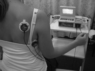
Additional biofeedback can be provided through the use of surface EMG, video and mirrors. Experience at our unit has found the use of surface EMG helpful in training appropriate activation patterns in shoulder movement (Malone et al 2006a) and has been shown to be more effective in regaining normal stability then strengthening exercises alone (Beall et al 1987, Reid et al 1996, Kiss et al 2001, Magarey & Jones 2003) (Fig. 12.1.5). Biofeedback techniques appear to facilitate the feed forward learning process at a cortical level, which may help to achieve more efficient motor patterns.
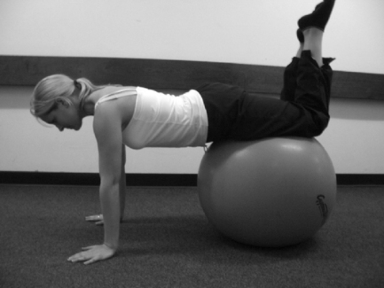
Weight-bearing exercises enhance joint stability, stimulate muscular co-activation and facilitate proprioception (Wilk et al 1993, Dines & Levinson 1995, Lephart & Henry 1996, Kibler 2000b). These can initially be done on a fixed base of support, progressing to stabilizing on a ball or balance board, enhancing neuromuscular control at a reflex level (Fig. 12.1.6). Strengthening exercises to the rotator cuff and deltoid can be introduced once the patient has achieved postural correction and the GHJ is located. The load applied must be graded appropriately to ensure that the correct muscles are strengthened. It is often unnecessary to use a Cliniband greater than yellow for cuff strengthening, where the emphasis should be placed on repetition rather than load to help build on endurance of the muscle.
Advanced stage
End-stage rehabilitation should focus on retraining patterns of movement biased towards functional tasks. Functional exercises, such as throwing, require co-ordination among multiple muscle groups, so it is important to address the entire kinetic chain (Dines & Levinson 1995, Kibler et al 2001). Repetition, speed and load may be varied in relation to the desired task, facilitating feed forward processing (Griffin & Letha 2003).
Proprioceptive neuromuscular facilitation (PNF) is useful to gain stability and control into functional patterns, strengthening through range (Voss et al 1953), while emphasis is placed on eccentric loading of the rotator cuff. It is important not to bombard the patient with numerous exercises, but to build on two or three sequentially. The frequency of exercise should be little-and-often, guided by fatigue and pain. Wherever possible, they should be made simple so they can easily be performed throughout the day. Patients who have particular problems with pain, will have to pace their activities, to gain a balance between appropriate use and exercise. The duration of management will vary amongst patients; on occasion, correction of a movement fault can be immediate, but it can take several months for the corrected pattern to become established at an unconscious level. It is also important to note that relapses may occur if a new task is undertaken, a growth spurt occurs, or the patient fails to adapt motor responses (Shumway-Cook & Woollacott 2001).
Summary
Important messages
Adib N., Davies K., Grahame R., et al. Joint Hypermobility syndrome in childhood. A not so benign multisystem disorder? Rheumatology. 2005;44:744-750.
Aronen J.G., Regan K. Decreasing the incidence of recurrence of first time anterior shoulder dislocations with rehabilitation. Am J Sports Med. 1984;12(4):283-291.
Barrett C., Emery R, Wallace A, et al. Altered corticospinal control of shoulder musculature in shoulder instability – a case study. Proceedings of the third conference of the International Shoulder group. Newcastle-upon-Tyne, Sept 4–6, 2000. 2000.
Beall M.S., Diefenbach G., Allen A. Electromyographic biofeedback in the treatment of voluntary posterior instability of the shoulder. Am J Sports Med. 1987;15(2):175-178.
Blasier R.B., Carpenter J.E., Huston I.J. Shoulder proprioception: Effects of joint laxity, joint position and direction of motion. Orthop Rev. 1994;23:45-50.
Burkhead W.Z., Rockwood C.A. Treatment of Instability of the Shoulder with an exercise program. J Bone Joint Surg Am. 1992;74:890-896.
Chu J.C., Kane E.J., Brent A.L., et al. The Effect of a Neoprene Shoulder Stabilizer on Active Joint-Reposition Sense in Subjects With Stable and Unstable Shoulders. Journal of Athletic Training. 2002;37(2):141-145.
DeCarlo M., Malone K., Gerig B., et al. Evaluation of Shoulder instability braces. Journal of Sports Rehabilitation. 1996;5:143-150.
Dines D.M., Levinson M. The Conservative Management of the Unstable Shoulder including Rehabilitation. Clin Sports Med. 1995;14(4):797-816.
Gibson J.C. Mini-Symposium: Shoulder Instability (iii) Rehabilitation after shoulder instability surgery. Current Orthopaedics. 2004;18:197-209.
Gibson J.C., Rayner V.S., Frostick S. Expanded Assessment in Multidirectional instability: An aid in planning effective rehabilitation?. 9th International Congress on the Surgery of the Shoulder, 2004. Washington USA, May 2004. 2004.
Griffin E., Letha Y. Neuromuscular Training and Injury Prevention in Sports. Clinic Orthopaedics and Related Research. 2003;409:53-60.
Hodges P.W., Cresswall A.G., Thorstensson A. Perturbed Upper Limb Movements Cause Short-Latency Postural Responses in Trunk Muscles. Exp Brain Res. 2001;138:243-250.
Ide J., Maeda S., Yamaga M., et al. Shoulder strengthening exercise with an orthosis for multidirectional shoulder instability: Quantitive evaluation of rotational shoulder strength before and after the exercise program. J Shoulder Elbow Surg. 2003;12(4):342-345.
Kendall F.P., McCreary E.K., Provance P.G., et al. Muscles: Testing and Function with Posture and Pain, ed 4. Baltimore: Williams & Wilkins, 1993.
Kibler W.B. Evaluation and Diagnosis of Scapulothoracic Problems in the Athlete. Sports Med Arthrosc. 2000;8:192-202.
Kibler W.B. Closed Kinetic Chain Rehabilitation for Sports Injuries. Phys Med Rehabil Clin N Am. 2000;11(2):369-384.
Kibler W.B., McMullen J., Uhl T. Shoulder rehabilitation strategies, guidelines and practice. Orthopaedic Clinics of North America. 2001;32(3):527-538.
Kiss J., Damrel D., Mackie A., et al. Non-operative treatment of multidirectional shoulder instability. International Orthopaedics (SICOT). 2001;24:354-357.
Kuroda S., Sumiyoshi T., Moriishi J., et al. The natural course of shoulder instability. J Shoulder Elbow Surg. 2001;10(2):100-104.
Lephart S.M., Henry T.J. The Physiological basis for open and closed kinetic chain rehabilitation for the upper extremity. Journal of Sports Rehabilitation. 1996;5:71-87.
Lephart S.M., Henry T.J. Restoration of proprioception and neuromuscular control of the unstable shoulder. In: Lephart S.M., Fu F.H., editors. Proprioception and Neuromuscular Control in Joint Stability. USA: Human Kinetics; 2000:405-413.
Lewis A., Kitamura T., Bayley J.I.L. Mini symposium: Shoulder instability (ii) The classification of shoulder instability: New light through old windows. Current Orthopaedics. 2004;18:97-108.
Magarey M.E., Jones M.A. Dynamic evaluation and early management of altered motor control around the shoulder complex. Man Ther. 2003;8(4):195-206.
Malone A.A., Jaggi A., Calvert P.T., et al. Muscle patterning instability – classification and prevalence in a reference shoulder service. In: Norris T.R., Zuckerman J.D., Warner J.J.P., Lee Q.T., editors. Surgery of the Shoulder and Elbow: An International Perspective. Rosemont, Illinois, USA: American Academy of Orthopaedic Surgeons, section 7, 2006.
Malone A.A., Noorani A., Jaggi A., et al. The role of dynamic electromyography in muscle patterning instability. Abstract proceedings, British Elbow & Shoulder Society 17th Annual Scientific Meeting, Edinburgh, 2006b. 2006.
McMullen J., Uhl T.L. A Kinetic Chain Approach for Shoulder rehabilitation. Journal of Athletic Training. 2000;35(3):329-337.
Mottram S.L. Dynamic Stability of the Scapula. Man Ther. 1997;2(3):123-131.
Myers J.B., Lephart S.M. Sensorimotor deficits contributing to glenohumeral instability. Clin Orthop Relat Res, 400. 2002:98-104.
Myers J.B., Wassinger C.A., Lephart S.M. Sensorimotor contribution to shoulder stability: effect of injury and rehabilitation. Man Ther. 2006;11:197-201.
Reid D.C., Saboe L.A., Chepeha J.C. Anterior Shoulder Instability in Athletes: Comparison of isokinetic resistance exercises and an electromyographic biofeedback re-education program – A pilot program. Physiotherapy Canada. 1996;48(4):251-256.
Shumway-Cook A., Woollacott M.H. Abnormal Postural Control. Motor Control Theory and Practical Applications, ed 2. Philadelphia: Lippincott Williams & Wilkins. 2001.
Takwale V.J., Calvert P., Rattue H. Involuntary positional instability of the shoulder in adolescents and young adults. J Bone Joint Surg. 2000;82B:719-723.
Thomas S.C., Matsen F.A. An approach to the repair of avulsion of the glenohumeral ligaments in the management of traumatic anterior glenohumeral stability. J Bone Joint Surg. 1989;71A:506-513.
Ulkar B., Kunduracioglu B., Cetin C., et al. Effect of positioning and bracing on passive position sense of shoulder joint. Br J Sports Med. 2004;38(5):549.
Voss D.E., Knott M., Kabat M. Application of Neuromuscular Facilitation in the Treatment of Shoulder Disabilities. Phys Ther Rev. 1953;33:536-541.
Wilk K.E., Arrigo C. Current Concepts in the Rehabilitation of the Athletic Shoulder. J Orthop Sports Phys Ther. 1993;18(1):365-378.
Zattara M., Bouisset S. Posturo-kinetic organization during the early phase of voluntary upper limb movement. 1 Normal Subjects. J Neurol Neurosurg Psychiatry. 1988;51:956-965.
12.2 The hand
Introduction
Hand dysfunction is commonly linked to ligamentous laxity (Murray 2006). The carpometacarpal joint (CMCJ) of the thumb may be particularly susceptible to weakened ligamentous constraints in the Ehlers-Danlos syndrome (Moore et al 1985). Symptoms may only present if the joint has been subject to excessive trauma, overuse or misuse and thus strains of the surrounding muscles and ligaments may have developed (Wynn Parry 2000).
Assessment of the hand
If appropriate and safe to do so, hand therapy patients are routinely assessed for hypermobility utilizing the 9-point Beighton score (Chapter 1). Other relevant joints are assessed using passive and active range of motion measurements, and appropriately recorded utilizing the standardized goniometry guidelines established by the American Society of Hand Therapists. It is important not to limit the assessment to the joints examined in the Beighton Scale, since the patient may be hypermobile in other joints. The scale is not all-encompassing and should be used only as a guide to the level of hypermobility displayed by an individual.
Circumferential measurements of joints using finger tape measures that are provided by Jobst™ are taken as appropriate. The modified Oxford Scale of manual muscle testing (Kendall et al 1993) is used to assess relevant muscle groups and as a way of assessing if the treatments are having an effect in increasing muscle strength and, in turn, joint stability. Maximal grip and pinch strength are measured using a hydraulic hand dynamometer and pinch meter, such as Jamar dynamometer and the pinch gauge by B&L Engineering (Mathiowetz 2002, Massy-Westropp et al 2004, Coldham et al 2006) and these again can map the patient’s progress and provide feedback as to the tolerance to exercises and functional activities.
As mentioned previously, hypermobility may only be present in one specific joint, and not global, and thus careful assessment is required in order to fully ascertain the patient’s symptoms and why the pain or functional difficulties are occurring. Assessment whilst performing functional tasks such as playing a musical instrument, writing or using cutlery is imperative, as hyperlaxity may be more evident whilst doing these activities (Fig. 12.2.1). It is important to distinguish between finger joint hyperextension, which can be a normal phenomenon, and lateral instability, which is often acquired or pathological (Tubiana 2000). Some level of hyperextension can be useful and assistive with certain task performance and the individual may have been born with a certain amount of hyperextension. If, however, the instability is acquired or pathological, the mechanisms and reasons why the phenomenon is occurring need to be analysed and addressed appropriately, in order to decrease progression of the condition.

Treatment principles
Symptom management is the key factor in this patient group. Initially, treatment may focus on decreasing an acute episode of pain through resting the affected area. Splinting is particularly important in this respect. In time, treatment must focus on joint stability, muscle strengthening, sensorimotor retraining to improve proprioception and patient education regarding healthy joint use (Warrington 2003).
The assessment and treatment of symptoms and dysfunction in the hand due to hypermobility should be considered in the context of the whole person. This will include attention to other areas of the body with symptoms, in particular the shoulder (Chapter 12.1) and cervical spine (Chapter 12.7), as well as considering postural alignment (Chapter 9) and general fitness (Chapter 13).
Joint protection and energy conservation
Hand therapists can assist in giving advice about joint protection and energy conservation techniques. These principles have been modified from those utilized for patients with rheumatoid arthritis (Boxes 12.2.1 and 12.2.2).
BOX 12.2.1 Methods of joint protection


5 Avoid activities that could lead to over-extension
Some directions of force can lead to over-extension at the joints.


BOX 12.2.2 Methods of energy conservation
1 Balance, rest and activity
 Try to plan ahead. Write a weekly or daily diary with activities in red and rest times in blue. Think about what you need to do and space the harder activities out over time.


Strengthening exercises
Patients can benefit greatly from a rehabilitation programme to improve joint control, muscle power (Wynn Parry 2003) and stamina (Wynn Parry 2000). Initially, stability exercises include isometric muscle contraction in a pain-free range (with a support on if being used) to encourage co-contraction of the muscles surrounding a joint. Involving the target object can be a useful progression to develop isometric strength and proprioceptive awareness, by maintaining the neutral joint position while holding a pen, violin bow or clarinet (Warrington 2003) and while performing the functional task. Later, exercises can be progressed to include concentric and eccentric strengthening.
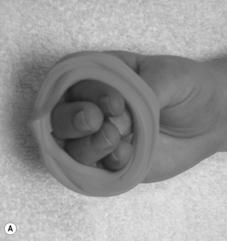
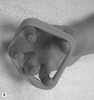
The intrinsic and extrinsic muscles of the hand are frequently stressed in an attempt to compensate for joint instability (Brandfonbrener 1990). Therapeutic putty exercises can be useful for specific muscle strengthening and in turn joint stability. Intrinsic muscle strength is very important and, because these muscles fatigue quickly, short pain-free sessions of exercise are efficient and encouraged (Davis & Rogers 1998). It is imperative that the exercises are performed with slightly flexed joints, rather than collapsing into hypermobile positions (Fig. 12.2.2). The use of graded rubber bands to assist in strengthening the interossei and lumbricals of the hands is also recommended (Wynn Parry 1998).
Strength gains are slower in the hypermobile patient, possibly due to alterations in central and peripheral neuromuscular physiological processes. It may therefore take many months for stability and strength to improve enough for the patient to be able to perform the task in a modified way and so a graded return to task performance is often required (Simmonds & Keer 2007). A diary of writing retraining times or musical practice schedule may be necessary to monitor symptoms and tolerable time of task performance. Exercises must be continued until sufficient muscle strength has been gained so a neutral joint position can be maintained whilst performing the required task.
Proprioceptive retraining
The product 3M™ Coban™ can be wrapped around a pencil, a bow of a stringed instrument, a stick of a drum or directly onto the finger to assist in retraining appropriate amounts of pressure applied when holding these items, and in turn facilitate an increase in proprioceptive awareness. This can lead to a decrease in the amount of muscle energy exerted to perform the task and thus fatigue and pain levels can be significantly decreased. Research performed by Lowell et al (2003) found that 3M™ Coban™ was effective in decreasing oedema in the skin-grafted burnt hand, and that this contributed to improved hand function, range of motion and strength levels with no impact on hand mobility, grip strength or function.
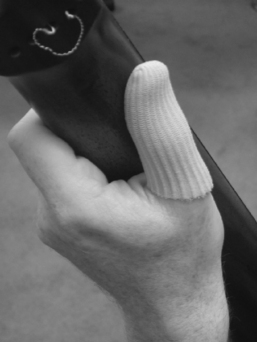
The use of a foam pad, lycra or silicon sleeve such as Silipos® can assist with proprioceptive retraining while performing a task such as playing a musical instrument (Fig. 12.2.3).
Splinting
Splints are a useful tool for supporting a joint in a neutral position to assist in decreasing joint strain and allow functional activities without pain. They are also thought to retrain proprioceptive awareness. Brandfonbrener (2003) comments that the use of ring splints for musicians with unstable fingers caused by ligamentous laxity helps prevent hyperextension but also helps retrain proprioceptivity in how they position their fingers.
Related posts:
 Anxiety disorders, their relationship to hypermobility and their management
 Fibromyalgia and hypermobility
 The physiology of pain
 What is the joint hypermobility syndrome?: JHS from the cradle to the grave
 Principles of rehabilitation and considerations for sport, performance and fitness
 Neuromuscular physiology in joint hypermobility
Stay updated, free articles. Join our Telegram channel

Full access? Get Clinical Tree


