CHAPTER 176
Pyogenic Granuloma or Lobular Capillary Hemangioma
(Proud Flesh)
Presentation
Often there is a history of a laceration or minor trauma to the skin or mucous membrane several days to a few weeks before presentation, but in most cases there is no apparent cause. The head, neck, and extremities are most commonly involved, especially the lips, oral mucosa, and fingers. An unsightly lesion forms, beginning as an extremely friable red or yellow papule or polyp, which now bleeds with every slight trauma. Objective findings usually include a crusted, sometimes purulent-appearing collection of erythematous, well-demarcated, red granulation tissue arising from a moist, sometimes hemorrhagic, skin ulceration, often with a collarette of scale at the base. There are usually no signs of a deep tissue infection (Figure 176-1).
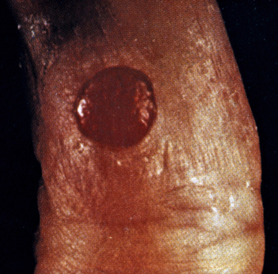
Figure 176-1 Pyogenic granuloma.
What To Do:
 Cleanse the area with an agent such as hydrogen peroxide or povidone-iodine solution.
 For small lesions (≤0.75 cm), cauterize the granulation tissue with a silver nitrate stick until it is completely discolored. Excess granulation tissue may first be shaved away using a No. 15 scalpel blade. The base of the lesion can be injected with 1% lidocaine with epinephrine to minimize the resulting hemorrhage. Thermal cautery may also be used, but local anesthesia will probably be required because of the potential inadvertent contact with intact dermis.
 Dress the wound after applying an antibacterial ointment, and have the patient gently wash and repeat ointment and dressings two to three times per day until healed.
 For pedunculated lesions, an atraumatic, simple, fast, and cost-effective alternative that does not require anesthesia is to ligate the base of the granuloma using a soft, absorbable, surgical suture material. This maneuver can be facilitated by lifting the pyogenic granuloma with forceps. The tissue is ligated with knots that are as tight as possible. The suture material is then snipped short. This can be covered by a simple wound dressing, and the tumor can be expected to become necrotic and fall off in several days. Inform patients or parents that although uncommon, bleeding could occur, and simple continuous compression for several minutes will control any minor hemorrhage. If the ligature does not reach far enough down to include the nurturing vessel, part of the granuloma may persist. This smaller lesion can then be easily treated with silver nitrate or thermal cautery.
 Patients with large lesions can be referred for treatment with a pulsed-dye laser.
 Warn the patient about the potential signs of developing infection and the need to return for treatment.
 Patients and parents should also be alerted to the possibility of recurrence after removal.
 Dermatology referral is recommended if the lesion recurs or multiple satellite lesions occur after excision.
What Not To Do:
 Do not cauterize or ligate any lesion that by history and appearance might be neoplastic in nature. Only treat clinically obvious cases. Pyogenic granuloma is occasionally confused with amelanotic melanoma, basal cell carcinoma, and squamous cell carcinoma. Periungual malignant melanoma can mimic pyogenic granuloma. Suspicious lesions should be referred for complete excision and pathologic examination.
 Do not cauterize a recurrent, large, or extensive lesion. These should also be considered for complete excision.
Discussion
Pyogenic granulomas, also known as lobular hemangiomas, are common vascular tumors of the skin and mucous membranes usually seen in children and young adults. They are usually smaller than 1 cm but can be up to 2 cm. Their cause is unknown, but pyogenic granuloma is a misnomer, because it is probably not infectious (pyogenic) in origin. It has been argued to be inflammatory and hyperplastic rather than a true neoplasm. Rapid growth is in response to an unknown stimulus that triggers endothelial proliferation and angiogenesis. They can be solitary or multiple and can arise within preexisting lesions, such as spider angioma and port wine stain.
Although a minority of pyogenic granulomas involute spontaneously within 6 months, most patients seek treatment because of bleeding. Pyogenic granulomas recur if any abnormal tissue remains.
Gingival lesions are common in pregnant women, in whom these lesions are called epulis gravidarum. Spontaneous resolution occurs after delivery.
It is not uncommon for secondary cellulitis to develop after the pyogenic granuloma is cauterized. It is therefore reasonable to place a patient on a short course (3 to 4 days) of a high-dose antibiotic (dicloxacillin or cephalexin, 500 mg qid, or cefadroxil, 500 mg bid) when the wound is located on a distal extremity.
Related posts:
Stay updated, free articles. Join our Telegram channel

Full access? Get Clinical Tree


