Fig. 32.1
Posterior cervical instrumentation and fusion of C1 ring fracture
In preparation for these surgical procedures, a thorough evaluation of the bony and ligamentous anatomy guides the surgical approach and technique. This evaluation is accomplished with standard radiologic evaluations (anterior, lateral, flexion, and extension views of the cervical spine), computed tomography (CT), and magnetic resonance imaging (MRI).
X-ray and CT scan are used primarily to evaluate bony abnormalities, while an MRI allows for evaluation of discoligamentous pathology and provides clear visualization of the spinal cord with any impingement or compression. If severe, spinal cord damage can be visualized with the MRI T2-weighted and STIR (short tau inversion recovery) images, spinal cord enhancement on the T2-weighted images may reflect pathologic changes due to inflammation, edema, ischemia, gliosis, or myelomalacia [1, 2] (Fig. 32.2). MRI with intravenous contrast can also help to assess blood flow as well as tumor margins.
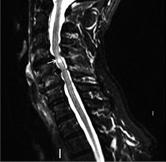
Fig. 32.2
MRI T2-weighted image with enhancing cord lesion (arrow) behind C5
Many surgical options have been described for posterior spinal fixation for fractures of C1 or C2, lateral mass/laminar fractures of C3–C7, or posterior decompression and stabilization for extensive spinal stenosis. These techniques include interspinous or sublaminar wiring, laminar hooks, lateral mass, and pedicle screw fixation [3]. Recent studies, however, have shown that three-column stability is best preserved with lateral mass and pedicle screw fixation because they provide (1) superior deformity correction, (2) greater immediate and long-term stabilization, and (3) can be used in almost all conditions requiring posterior stabilization or reconstruction of the occipitocervical spine, midcervical spine, or cervicothoracic junction [4, 5] (Fig. 32.3). Table 32.1 reviews the major indications for instrumentation with pedicle/lateral mass screws.
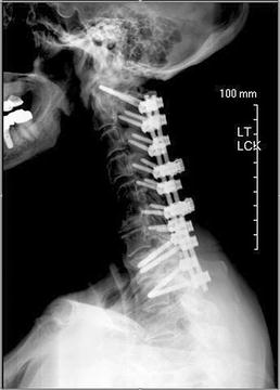
Fig. 32.3
Posterior laminectomy and fusion with pedicle screw instrumentation, C1, T1–T2 with lateral mass screw instrumentation C2, C3, C4, C5, C6, and C7
Table 32.1
Cervical pedicle /lateral mass screw indications
Posterior disruption or anterior/posterior disruption without severe vertebral body injury |
Cervical spinal instability caused by nontraumatic lesions (metastatic tumor, rheumatoid arthritis, and destructive infectious lesions) |
Correction of cervical malalignment in the sagittal plane, including postlaminectomy and posttraumatic kyphosis spondyloarthropathy |
Stabilization of the segmental motion caused by posterior decompression |
Posterior reduction and stabilization at the cervicothoracic junction |
Salvage of previous anterior surgeries |
Rigid stabilization of the craniocervical fixation |
In comparison to lateral mass screws, pedicle screws have a higher pullout force and the greatest capacity for restoring sagittal alignment of the cervical spine with correction of malalignment in the occipitoatlantoaxial region [6–8]. However, because of the size of the pedicle screws in relation to the pedicle and the narrow trajectory required, they also have a greater chance of nerve root or vertebral artery injury. Since the risks of neurovascular complications caused by malaligned screw placement into the cervical pedicle cannot be completely avoided, they are often placed in the higher cervical and cervical-thoracic levels where the pedicle has sufficient size for screw placement [9–14].
Cervical spinal myelopathy (CSM) is another common indication for a posterior cervical surgical procedure. In contrast to cervical fractures, which occur mostly in males younger than 40 years old, CSM is the most common cause of progressive neurologic decline in patients more than 50 years old [15, 16]. In most cases, myelopathy develops slowly due to spinal cord compression from progressive degenerative arthritis (osteophytes), ossification of the posterior longitudinal ligament (OPLL) or hypertrophy of the ligamentum flavum. The lower extremities are usually affected first with patients presenting with gait disturbances caused by the degeneration of the spinocerebellar and corticospinal tracts. Further compression or acute injury can result in upper extremities, loss of coordination, and difficulty with fine motor tasks [15].
In general, CSM most commonly affects the C5–C7 region of the cord with a variable clinical presentation. Often patients will have neck pain and stiffness and can experience deep aching or burning in the upper extremities (brachialgia). Motor and sensory dysfunction may be unilateral or bilateral depending on the extent and location of cord compression.
Patients with mild signs and symptoms of CSM are usually followed clinically. However, surgical decompression from an anterior or posterior approach is indicated in patients with progressive moderate to severe neurologic deficits. While the optimal surgical strategy (anterior vs. posterior or a combined procedure) for CSM) remains controversial, there is no clear evidence that either approach more reliably results in recovery from a compressive myelopathy [3, 14, 17] (Fig. 32.4). The goals of the surgical procedure are to decompress the spinal cord to avoid either static or dynamic compression, restore sagittal alignment, stabilize the spinal column, and avoid kyphosis. While the surgical procedure itself inconsistently improves myelopathic symptoms, it does prevent further decline of neurologic function [18, 19]. Thus, in many cases the surgery is prophylactic to prevent continued spinal cord damage and loss of function.
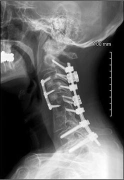
Fig. 32.4
Combined anterior (C3–C5) and posterior fusion with lateral mass screws, C2, C3, C4, C5, C7, and bilateral pedicle screws, T1 and T2
Because the majority of the abnormal anatomy-producing spinal cord compression is located anteriorly, the procedure most commonly chosen is an anterior cervical discectomy/corpectomy and fusion (ACDF; see Chap. 31). However, in multilevel compression and/or progressive deformity, posterior decompression with instrumentation may be indicated, often in combination with an anterior procedure. In the past, posterior decompressions alone often led to progressive kyphosis. With modern fixation techniques and the increased use of cervical instrumentation and fusion, this is much less common [4] (Fig. 32.5).
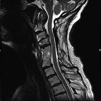
Fig. 32.5
Previous anterior cervical fusion at C4–5 with posterior laminectomy C3–5. The patient now presents for posterior cervical instrumentation secondary to spondylolisthesis at C2–4 and progressive kyphosis
With improvements in posterior instrumentation (screw and rod fixation) in the past decade, there has been an increased use of posterior decompression and instrumentation for the treatment of extensive degenerative cervical myelopathy. One complication of the extensive laminectomy is the formation of a thick fibrous scar at the operative site that replaces the bony compression and with progressive kyphosis reproduces the original symptoms in the extended postoperative period. An alternative technique to a laminectomy is a laminoplasty, which provides for relief of the pressure on the spinal cord with surgical reconstruction of the posterior vertebral elements to increase space for the neural structures while maintaining aspects of the bony posterior arch [20]. Thus, muscle reattachment is also possible to help maintain a posterior tension band and reduce the risk of postoperative kyphosis. A laminoplasty involves cutting through the lamina of the vertebrae on one side and merely cutting a groove on the other side and then “swinging” the freed flap of bone open to relieve the pressure on the spinal cord. The spinous process may be removed to facilitate the process. The bone flap is then propped open using small wedges or pieces of bone such that the enlarged spinal canal will remain in place [3, 21, 22] (Fig. 32.6). Mini-plates can also be used to maintain the open canal and increase stability.
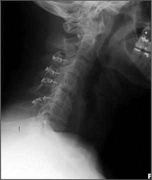
Fig. 32.6
Posterior cervical laminoplasty C3–C6
While the progression of CSM is highly variable, major neurologic injury from even relatively minor spinal cord injuries can manifest as one of four clinical syndromes (central cord syndrome, anterior cord syndrome, Brown-Sequard syndrome, and a transverse lesion syndrome). The most common location for spinal injuries from trauma is in the flexible regions of the cervical spine (e.g., C4–C5). The central vascular region of the spinal cord is the most easily injured, with progressive degrees of injury extending the injury radially until the entire cord is injured (transverse lesion syndrome). Anatomic knowledge of the spinal pathways of the syndromes helps guide the approach to interpretation of intraoperative neurophysiologic monitoring during posterior cervical decompression (Table 32.2).
Table 32.2
Cervical spinal cord injury syndromes —clinical findings and anatomic correlation
Syndrome | Clinical findings | Anatomy |
|---|---|---|
Central cord syndrome | Injury to central grey matter (anterior horn cells) or lateral columns with greater weakness or paralysis, pain and temperature loss of the upper extremities compared to the lower extremities | 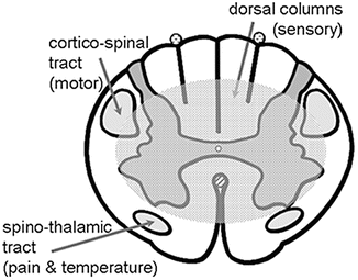 |
Light touch and proprioception is usually spared | ||
Brown-Sequard syndrome | Injury to the corticospinal tract, dorsal column, and spinothalamic tract resulting in: | 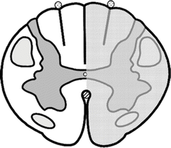 |
Ipsilateral weakness or paralysis | ||
Ipsilateral loss of proprioception and light touch loss | ||
Contralateral pain and temperature loss below the lesion | ||
Anterior cord syndrome | Loss of motor, pain and temperature, with preservation of fine touch and proprioception | 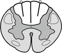 |
Transverse lesion syndrome | Loss of all function below the level of injury | 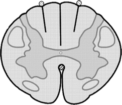 |
During posterior cervical procedures , the application of somatosensory- and motor-evoked responses is frequently helpful in the evaluation of intraoperative compromise of the spinal cord during the procedure and during positioning of the patient. While the use of intraoperative neuronavigation systems and fluoroscopy aids in placement of the pedicle screws, cortical perforation still occurs in 20–25 % of pedicle screw placements [11, 14]. A recent small study (n = 26) evaluated the sensitivity and specificity of pedicle/lateral mass screw stimulation thresholds for the determination of cortical perforation [23]. A stimulation threshold of 15 mA or greater provided a 99 % positive predictive value (89 % sensitivity, 87 % specificity) that the screw was within the lateral mass or pedicle. However, stimulation values of 10–15 mA provided an intermediate sensitivity and specificity (66 and 90 %, respectively); whereas a stimulation value of less than 10 mA was highly predictive that a screw is malpositioned (70 % sensitivity, 100 % specificity). Thus, the addition of the intraoperative evoked EMG monitoring is a valuable tool in the evaluation of lateral mass and pedicle screw placements with stimulation values below 10 mA triggering evaluation, repositioning, and possible removal of the pedicle screw.
Related posts:
 Deep Brain Stimulation
 Intraoperative Neurophysiologic Monitoring During Surgery for Supratentorial Mass Lesions
 Surgery for Chiari Type I Malformation
 Intraoperative Neurophysiological Monitoring for Intracranial Aneurysm Surgery
 Trigeminal Microvascular Decompression
 Monitoring of Jugular Venous Oxygen Saturation
Stay updated, free articles. Join our Telegram channel

Full access? Get Clinical Tree


