The popliteal crease is identified and marked while the patient flexes the knee. The medial and lateral borders of popliteal fossa are formed by semimembranosus and biceps femoris tendons. The sciatic nerve is located between these two muscle groups.
Approach and Technique
 With patient in prone position, a high frequency (HF) linear transducer is oriented perpendicular to the long axis of the femur over the intertendinous junction.
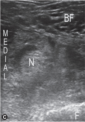
 Identify the sciatic nerve in short axis anterior and medial to the fascia of biceps femoris muscle, posterior and medial to femur, and posterior to the popliteal vessels when visible.
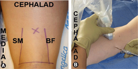
A. The popliteal fossa is bordered laterally by the biceps femoris tendon and medially by the tendons of semimembranosus and semitendinosus (right popliteal fossa shown). The sciatic nerve is reliably located below the intertendinous junction identified by the “X.” B. The ultrasound transducer is oriented perpendicular to the long axis of the femur across the intertendinous junction to image the sciatic nerve in cross-section. The block needle is inserted lateral to the transducer and directed medially for in-plane needle guidance (left thigh shown).
US-GUIDED POPLITEAL FOSSA SCIATIC BLOCK
Related posts:


Stay updated, free articles. Join our Telegram channel

Full access? Get Clinical Tree


