Fig. 18.1
“Free Air” is seen here under the right diaphragm. © Dale Dangleben, MD
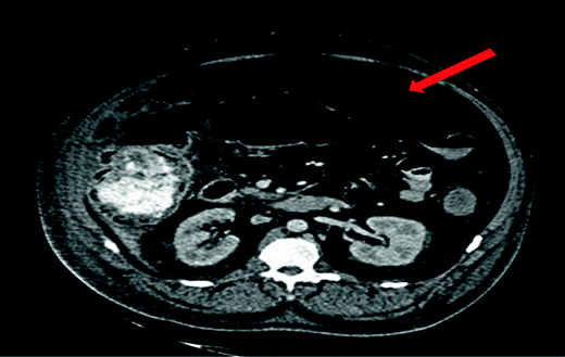
When plain radiographs are non-diagnostic and the patient is stable and able to undergo additional imaging studies, computed tomography (CT) is the next study of choice for adult patients as it is a more sensitive study. A CT scan may also help to localize the site of perforation and hence aid in operative planning but should not delay surgical intervention in an acutely ill patient (Fig.18.2).

Fig. 18.2
Large amount of pneumoperitoneum “Free Air” is seen here outside the colon. There is also pneumatosis of the right colon. © Dale Dangleben, MD
In children or pregnant patients, one should consider ultrasonography, as in some cases ultrasound may be superior to plain films in these groups.
One should also be aware of “pseudopneumoperitoneum,” or air in the abdominal cavity that appears to be intraperitoneal but is actually contained in an organ. An example of such a finding is Chilaiditi’s sign: colonic interposition between the liver and diaphragm [1].
Complications
Complications of pneumoperitoneum due to perforated viscus include abscess or phlegmon formation, peritonitis, septic shock, multi-organ failure and death should the diagnosis be delayed or missed. Early intervention is crucial, as patients with perforated viscus and peritonitis have a mortality rate of greater than 20%.
Stay updated, free articles. Join our Telegram channel

Full access? Get Clinical Tree


