Issue to address
Method of investigation
Spinal anatomy for needle placement
Visual inspection of the spine
Spinal anatomy for needle placement
Plain films of the lumbar spine
Ability to thread catheter
Review previous procedures
Ability to thread catheter
MRI or CT if history of complex anatomical anatomy
Level of needle placement
Plain films of the lumbar spine and inspection of skin
Course of tunneling
Visual inspection of the patient’s body
Pocket placement
Drawing of planned pocketing including palpation of rib and anterior iliac spine
Fig. 37.1
Lateral decubitus positioning of patient (posterior view (a) and anterior view (b))
In some cases the preoperative evaluation will suggest that there is not an appropriate area to place a pump in the abdominal wall. Reasons may include lack of body fat, previous surgeries, or skin infection or poor quality of skin texture.
An alternative position is prone placement of the pump, which obviates the need for lateral decubitus position. In the lateral decubitus position, the patient should be aligned with both shoulders at equal tilt to avoid torsion of the trunk. The patient is prepared widely to encompass both the planned area of invasion and the surrounding tissues. Fluoroscopic imagery is used to assess the bony anatomy and to determine the level in which the needle will be placed. Once the clinician is pleased with the positioning, preparing, draping, and x-ray imaging, the procedure is initiated. In some cases the position is less than optimal because of the patient’s pain being too great to be placed in the lateral decubitus position or because of body habitus and spinal abnormalities. In these cases the position can be modified to the prone orientation, realizing that, if the plan is to place the pocket into the abdominal wall, repositioning will be required. However, if there is adequate body mass to support the pump in the posterior flank or above the hip, then pump placement in the buttock or hip area may be considered, thus avoiding the need to reposition the patient during the procedure. It is important to realize that the prone position buttock placement of the pump should be used with pumps of a volume of 20 mL or less, given their inherent bulk when compared with neurostimulator implanted pulse generators. In addition, the pump should be placed at least 10 cm above the iliac crest to avoid uncomfortable pressure on the bony surface when the patient is sitting or lying down.
37.2.1 Needle Placement
After satisfactory positioning, preparing, and draping, the fluoroscopic image is visualized to place local anesthetic one to two vertebral bodies below the planned site of entry. A paramedian approach is recommended to allow the catheter to avoid the constant wear and tear of the spinous processes (Fig. 37.2). The needle is positioned to walk off the lamina and enter the intrathecal space at an ideal angle of 30° to 45° (Fig. 37.3). In some cases the angle can be must be increased to 60° because of anatomical variation, but angles that exceed this mark may lead to excessive pressure on the catheter and subsequent catheter failure (Fig. 37.4). Another approach to imaging needle guidance includes the use of the contralateral oblique view (CLO). In the CLO view the image intensifier is inserted obliquely up to 45° away from the clinician (Fig. 37.5). The needle, in many cases, can be seen piercing the ligamentum flavum to enter the epidural space and then into the intrathecal space (Fig. 37.6).
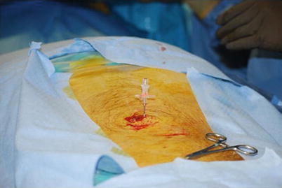
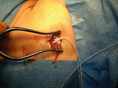
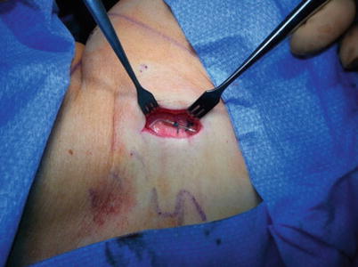
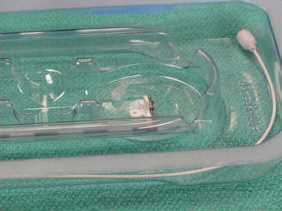
Fig. 37.2
Paramedian approach to needle placement
Fig. 37.3
Proper needle angle placement at 30°
Fig. 37.4
Placement of anchor before catheter tunneling
Fig. 37.5
Placement of anchors and stress relief loop, 2 catheter technique
Fig. 37.6
Example of an intrathecal catheter with stylet, needle, anchor, and titanium connector
Once the needle is positioned into the intrathecal space, the implanter should remove the stylet and visualize unobstructed flow of cerebrospinal fluid (CSF). In some settings physicians have attributed lack of flow to scarring or narrowing in the canal. No documented publication shows that a successful placement can be achieved with poor or no flow. In the setting of poor flow, the implanter should be suspicious of epidural or other placement outside of the CSF. The authors would recommend repositioning the catheter until flow is satisfactory. Once good flow of CSF is confirmed, the implanter can move forward with catheter placement.
Modern intrathecal catheters have improved steering ability and are more radiopaque. Ideally the catheter is placed in the posterior intrathecal space. The low angle of needle placement is important in this process. The level of tip placement varies on physician preference, but recent science suggests placement of the tip closest to the spinal cord level matching the dermatomal level of pain provides for improved chance of a satisfactory outcome. Some clinicians prefer to place the catheter below the conus to reduce the complication severity of a granuloma, but this has not been proven to be accurate. The improved position may be less helpful in pain relief and necessitate increased drug dosing, which may negate the benefit of placing the catheter below the conus.
Related posts:


Stay updated, free articles. Join our Telegram channel

Full access? Get Clinical Tree


