Age group
RR (breaths per min)
HR (beats per min)
BP (systolic; mmHg)
Newborn (birth to 28 days)
40
140
60
Infant (under 1 year)
30
120
80
Child (approx 3–6 years)
20
100
100
Adolescent
Teens
80
120
High metabolic rates associated with small size, demands of growth, and combating heat loss also account for high resting heart rates in the newborn (140/min) and infant age groups (120). The small size of infant hearts limit stroke volume, so any adjustments in cardiac output must come from changes in heart rate. Bradycardia (under 100 beats per minute) in infants is always ominous. Heart rates below 100 usually are due to hypoxemia and demand immediate attention. Supplemental oxygen must be administered and if the heart rate remains low external cardiac massage is mandatory. Small children have resting rates of 100, a number that may be concerning to practitioners used to slower adult values typical of adolescents (under 80).
Normal blood pressures among newborns (60 mmHg systolic) and infants (80) reflect the limited cardiac performance of the infant heart. A corollary is that any decrease in systolic blood pressure is a preterminal sign and immediate measures must be taken to support the blood pressure either with volume, pharmacological intervention, or external cardiac massage. Normal blood pressures increase with older children (100) and reach adult values in adolescents (120).
Body temperature, however, is the same in all age groups, 37 °C. In newborns and infants a skin temperature from the axilla is routine, and a slightly lower value expected (36.0–36.5°). Oral temperatures are hard to obtain and rectal values unnecessary (and potentially dangerous). Newborns and infants are especially prone to hypothermia because they have large surface area relative to body size, depend on brown fat metabolism for thermogenesis, and are unable to shiver to generate heat. Young patients should be wrapped in warm blankets before and after being seen and placed under warming blankets and radiant heat lamps when being examined or receiving active therapy.
Any deviation from normal values requires constant electrocardiographic and respiratory monitoring, pulse oximetery, and periodic measurement of blood pressure. Admission or transfer to a higher level of care is required for persistent tachycardia or tachypnea, periodic breathing (periods of cessation of breathing efforts lasting seconds and not associated with other changes in heart rate), apnea (no breathing effort of 15 s or more, or any cessation of breathing effort associated with bradycardia), bradycardia, low blood pressure, and change in sensorium (persistent agitation, irritability, restlessness, stupor, or coma).
Airway and Breathing
Signs that an infant has respiratory distress can be subtle but demand the administration of supplemental oxygen and transfer to a higher level of care. Infants exhibit tachypnea, pallor, nasal flaring, and air hunger. Hypoxemia may become manifest as irritability and restlessness, so caution demands an evaluation with a pulse oximetry before sedatives or analgesics are considered. Retractions of the chest wall and soft tissues of the lower neck and upper chest reflect increased work of breathing. As the infant tires or begins to decompensate periods of apnea occur. Bradycardia, a late sign, indicates hypoxemia and impending cardiac arrest. Cyanosis is also a late sign and may not be seen because the patient may decompensate before desaturated hemoglobin levels reach 5 g/dL, the concentration associated with the color.
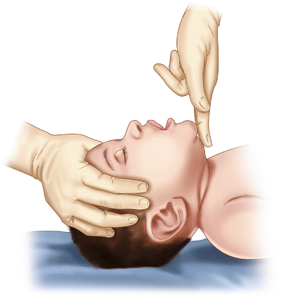
Infant airways tend to obstruct. Their large head and prominent occiput flex the neck and collapse the upper airway, easily opened by head tilt and jaw lift maneuvers (Fig. 34.1). Any parent knows that young children always have food and a variety of other foreign material near or in their mouths, always are eating, and frequently have a respiratory infection. Suction equipment must be close at hand. Early placement of a nasogastric tube is a necessary step in airway control to decompress the stomach and prevent aspiration. A common error is using a feeding tube that is too small for adequate decompression. Infants require at least a 10 French tube.


Fig. 34.1
Head tilt and chin lift in an infant. These are the first maneuvers to open the airway in infants and small children
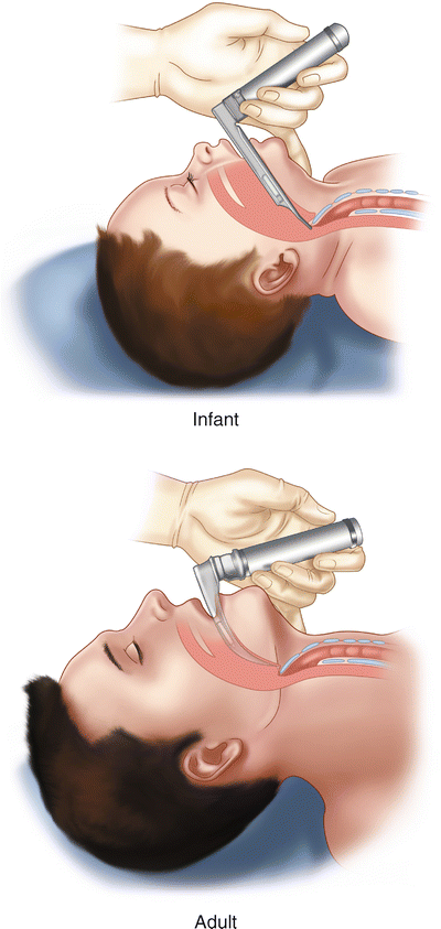
Supplies necessary for airway control must be at hand when seeing children with emergency conditions, including laryngoscopes and tubes of sizes appropriate for infants and small children. A useful memory aid is that the patient’s little finger approximates the appropriate size of the endotracheal tube. Supplies must be inventoried and checked regularly to assure they remain in working order, especially items like laryngoscopes that have batteries and require illumination. Practice dummies of the infant head and neck allow practice intubation and fairly accurately reproduce the anatomic features that make the procedure a challenge. The orifice itself is small. The larynx lies high in the neck, obscured by the epiglottis, which is rigid and shaped like an “omega” rather than a broad based “U.” The maneuver to visualize the larynx is to lift the tip of the epiglottis with the tip of a straight laryngoscope blade (Fig. 34.2). If the larynx cannot be seen the usual error is that the laryngoscope is too distally placed.


Fig. 34.2
The view at direct laryngoscopy in an infant (left) and an adult (right). The tip of a straight laryngoscope blade lifts the epiglottis in the infant, while a curved scope used in adults pulls the vallecula, anterior to the epiglottis upward to visualize the adult larynx
A surgical airway may be mandatory in massive upper airway injuries where there is bleeding or soft tissue trauma. If the patient is exchanging air the best initial maneuver is to provide supplemental oxygen and move to the operating room where light, equipment, and trained assistance are at hand. The most experienced anesthesiologist or anesthetist then attempts orotracheal intubation without paralytics or anesthesia. If standard airway control is unsuccessful after multiple attempts then the most experienced surgeon performs a tracheotomy using an incision with which he or she is most comfortable. In such a situation the incision must be of adequate size to provide immediate access to the trachea and control bleeding. Use a standard pediatric endotracheal tube—in such a situation a standard tracheotomy appliance is too short and the stay collars are cumbersome.
Stay updated, free articles. Join our Telegram channel

Full access? Get Clinical Tree


