Percutaneous Neuromodulation Therapy

16 Percutaneous Neuromodulation Therapy


After using his own version of acupuncture for several years to treat complaints ranging from migraine headache to alcohol withdrawal to cancer pain, Craig decided in the late 1970s that he would develop a new system based on his belief that the effectiveness of electroacupuncture depended on its influence on the central nervous system. The new system was to be based on placing needles into the vicinity of nerves that conducted impulses away from the affected areas. The earliest recorded workshops called his technique “dermatomal electroacupuncture”.


The system evolved through the years as Craig manipulated the key components of treatment, fine-tuning such important aspects as needle location, overall montage of needles employed, and stimulus frequency and duration. In parallel, other scientists advanced our understanding of the neurophysiology of chronic pain, elucidating the role of various neurotransmitters and the impact of electrical stimulation on the behavior of various nerve pathways. By the mid-1990s, dermatomal electroacupuncture had become percutaneous electrical nerve stimulation (PENS), which soon became Craig-PENS to honor Craig’s contribution. Besides indicating what the technique involved (percutaneous stimulation of nerves with electrical impulses), PENS emphasized the demarcation of this technique from acupuncture. Many practitioners continue to use PENS or Craig-PENS to describe what they do.


A new millennium demanded a new acronym. At the turn of the century, Craig and colleagues renamed the technique Percutaneous Neuromodulation Therapy (PNT). This new moniker moved beyond description to explanation. PENS defines the technical performance of the method, but PNT explicates the neurophysiology behind why it works. Moreover, a new term was needed to distinguish an effective chronic pain treatment from a neurosurgical procedure involving implantable stimulating electrodes. This chapter will discuss PNT as developed by Craig and his associates.


A word about acupuncture: although Craig studied acupuncture and initially called his technique dermatomal electroacupuncture, PNT should be distinguished from electroacupuncture. Unlike electroacupuncture, PNT does not involve needling of specific, traditional acupuncture points. In fact, as will be described later, placement of needles in PNT intends to influence dermatomal (or myotomal or sclerotomal) nerve pathways rather than acupuncture meridians. And PNT does not rely on the concept of “qi” or require any sort of traditional Chinese medicine (TCM) diagnosis. However, like electroacupuncture, PNT does involve needle insertion and electrostimulation, so the results of neurophysiologic studies of electroacupuncture also reflect the neuromodulatory impact of PNT. The importance of differentiating PNT from electroacupuncture lies here: one need not undertake the study of acupuncture to to understand and practice PNT effectively.


How PNT Works


As its name suggests, PNT is believed to work by modulation of central pain pathways using peripheral nerve stimulation. A body of neurophysiology research supports this presumption, and ongoing work continues to reveal details of the complex interactions that underlie the perception of pain.


Peripheral nerve endings known as nociceptors sense pain and transmit signals to the dorsal root ganglion of the spinal cord. Localized, sharp, stinging pain, generally of short duration, reaches the cord by way of thicker, myelinated A-delta fibers. Dull, aching, burning pain, longer in duration and slower in onset, travels along smaller, nonmyelinated C fibers. When the pain signal reaches the dorsal horn, various neurotransmitters activate spinal neurons that send axons across the spinal cord and up fiber tracts that eventually reach the medulla, midbrain, and thalamus. From the thalamus, neurons project to the prefrontal cortex, the motor cortex, the somatosensory cortex, the limbic system, and the hypothalamus. Pain information also finds its way to the pons, cerebellum, and basal ganglia through pathways that remain to be fully elucidated.


Besides the modulation that can be brought about during the ascent of pain signals, descending inhibitory pathways activated in the midbrain, brainstem, and numerous other areas (among them the amygdala, the hypothalamus, the arcuate nucleus, and the periaqueductal gray matter) can modify the transmission of these impulses. PNT aims to influence the balance within this very complex network, enhancing the inhibitory drives and suppressing the excitatory message.


The transmission of pain signals throughout these pathways depends on the balance of neurotransmitters whose release depends, in turn, on a variety of factors, including the frequency of stimulation of the peripheral nerves. Rather than reviewing the various forms of gate theory, originally proposed by Melzack and Wall,1 we will focus here on the influence of different frequencies of electrostimulation on neurotransmitter release in the central nervous system because this neuromodulation underlies the therapeutic effectiveness of PNT. Much of this knowledge derives from electroacupuncture and neurosurgery research, as noted earlier.


Early work by Han and colleagues from Beijing Medical University demonstrated that analgesia induced by electroacupuncture of different frequencies is mediated by different opioid receptors.2 Analgesia induced by low-frequency (2 Hz) electroacupuncture resulted from activation of mu- and delta-opioid receptors, whereas analgesia induced by high-frequency (100 Hz) electroacupuncture derived from activation of kappa-opioid receptors. Intermediate frequency stimulation (2 to 15 Hz) activated all three types of receptors in the spinal cord of rats.


Further work by Han and colleagues3 showed that low-frequency (2 Hz) peripheral stimulation produces a significant increase in enkephalin release into the lumbar cerebrospinal fluid (CSF), whereas high-frequency (100 Hz) peripheral stimulation increases dynorphin release. This validated earlier work by Mayer and colleagues4 and Pomeranz and Chiu.5 Wang and associates went on to establish that a more potent analgesia could be established by asynchronous electroacupuncture stimulation—alternating between low-frequency (2 Hz) and high-frequency (100 Hz) stimulation—than could be attained by synchronous electroacupuncture stimulation, combining low and high-frequency stimulation at the same time.6 The clinical application of these findings will become evident later in this chapter.


The endorphins (enkephalin and dynorphin) are only part of an ever-elaborating story of pain neurotransmission. In the last few years, research has suggested a role in electroacupuncture (and, thus, PNT) for cocaine and amphetamine-regulated transcript (CART) peptide,7 arginine vasopressin,8 serotonin, catecholamine, and spinal Fos expression,9 interleukin-1-beta,10 corticotrophin-releasing hormone.11 No doubt more neurotransmitters and neurochemicals will play important parts in the transmission and perception of pain.


In summary, pain perception is complex and incompletely understood. How PNT modulates the perception of pain remains unclear. Nevertheless, based on this knowledge from basic research and the principles realized through careful clinical study, PNT can be used effectively to address a broad range of chronic painful conditions.


The PNT Technique


In a typical treatment session, ten 32-gauge stainless steel acupuncture needles are inserted to a depth of 2 to 4 cm into the soft tissues and/or paraspinous muscles in and around the dermatomal distribution that covers the painful site(s). The 10 needle probes are then connected to five bipolar leads, with each lead connected to one positive and one negative probe in a pattern designed to direct the stimulus along the path of the nerve supplying the dermatome. The probes are then stimulated at an alternating frequency of 15 Hz and 30 Hz for 30 minutes, with the intensity of the stimulation adjusted to produce a gentle tapping sensation without muscle contraction. Treatment sessions can occur once to several times per week. In published trials, treatments have ranged from 2 to 12 weeks, but the overall duration of treatment in the real clinical setting depends on the patient’s response.


The details of the typical treatment session have been defined through a number of clinical trials investigating specific aspects of the treatment. Here we will describe research supporting the location of the needle probes, the stimulation montage, the stimulation frequency used, and the duration of electrical stimulation.


Location of Needles


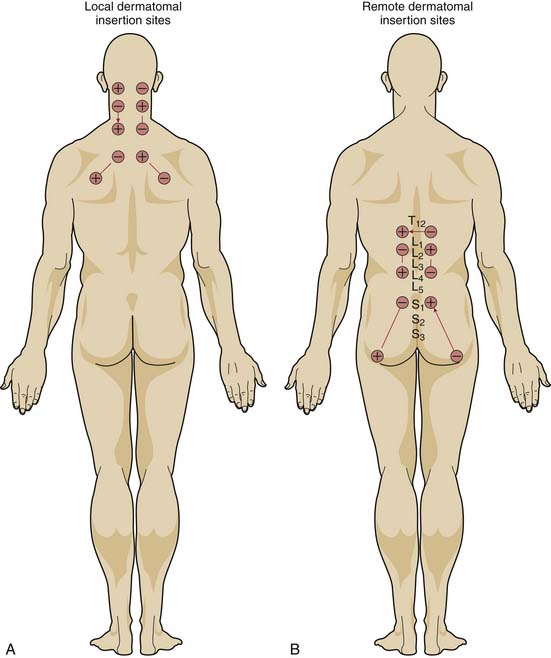
If the mechanism for analgesia relies primarily on increases in analgesic-like neurotransmitters within the central nervous system induced by peripheral nerve stimulation, varying the level of spinal stimulation should yield similar analgesic effects. Alternatively, if central neuromodulatory changes are primarily responsible for the analgesic effects, then stimulation of peripheral nerves in or near the affected area should prove more effective than stimulation of distant peripheral nerves. White, Craig, and others clarified the effect of the location of electrical stimulation on the acute analgesic response to PNT in a crossover study of 68 patients with nonradiating neck pain.12


When PNT was applied to needles located in the dermatomal distribution of the neck pain, visual analog pain scores decreased significantly more than when PNT was applied to needles located in the low back region or when needles were inserted without electrical stimulation. PNT applied to local needles also brought significant improvements in physical activity, quality of sleep, psychological well-being, and the need for oral analgesic medications, compared with PNT applied to distant needles or unstimulated needles. Moreover, unlike the other two treatments, PNT with the local needles showed a cumulative improvement over the course of therapy (Fig. 16-1).


At least in patients with neck pain, therefore, electrical stimulation at the dermatomal levels corresponding to the local pathology provides a greater analgesic effect than does stimulation at remote dermatomes, and electrical stimulation is required to provide the optimal hypoalgesic effect. In order for PNT to be most effective, the practitioner must identify the dermatomes that overlap and surround the painful area and place the needle probes within these dermatomes.


Stimulation Montage


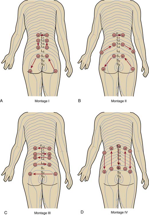
Having placed the needles into the appropriate dermatomes, does it matter how one connects the stimulator leads? In other words, does one specific pattern of electrical stimulation (i.e., montage) provide greater relief than another specific pattern? Work by White and associates demonstrates that montage matters.13


The investigators evaluated the effect of four different montages on the acute analgesic response to PNT when applied at the same dermatomal levels in a crossover study of 72 patients with low back pain (Fig. 16-2). Although all four montages provided significant improvements in pain visual analog scores and in the physical component summary (PCS) and the mental component summary (MCS) of the SF-36, one montage—in which the flow of electrical stimulation paralleled the dermatomes on both sides—produced significantly greater improvements. The cumulative effects over the course of 2 weeks of treatment were also superior with this montage than with the other patterns.


The researchers speculated that the stimulation pattern used with this montage produced more effective electrical stimulation of the involved myotomes and/or sclerotomes and concluded that this montage should be employed in the initial treatment. If this pattern fails, however, they advised resorting to another montage. When initiating PNT, therefore, the best montage employs needles within and around the affected dermatome and electrical stimulation bilaterally between needles in the same dermatome.


Jun 19, 2016 | Posted by in PAIN MEDICINE | Comments Off on Percutaneous Neuromodulation Therapy

Full access? Get Clinical Tree

Get Clinical Tree app for offline access