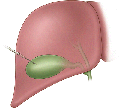
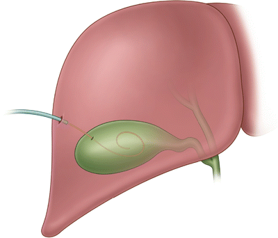
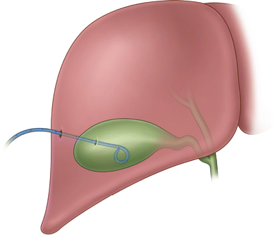
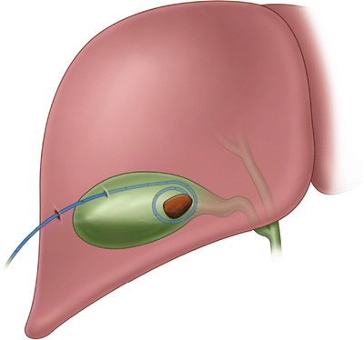
Fig. 6.1
Difference in approach for transhepatic versus transperitoneal approaches
Under real-time ultrasound guidance, the 21-gauge needle is advanced through the hepatic parenchyma into the gallbladder. Aspiration of either bile or purulent aspirate confirms proper position (Fig. 6.2).


Fig. 6.2
Needle entering gallbladder lumen as seen under fluoroscopy. Contrast can be appreciated in the dependent portion of the gallbladder
The syringe is then removed from the access needle and soft J-tipped guidewire inserted into the gallbladder. Insertion of the 0.018 guidewire into the gallbladder cavity should be confirmed with ultrasound guidance (Fig. 6.3). Access needle is removed and a 6 Fr sheath advanced over the wire. With the six sheath in place, the 0.018 is replaced with a 0.035 guidewire.


Fig. 6.3
Guidewire in gallbladder lumen as seen under fluoroscopy
The sheath is removed and an appropriately sized dilator (dependent on ultimate size of final catheter) is placed over the guidewire. The dilator is then removed; pigtail catheter inserted over the guidewire; the guidewire is removed leaving the catheter in position (Fig. 6.4).


Fig. 6.4
Pigtail catheter in final position within gallbladder lumen. Contrast can be appreciated filling the gallbladder
Again aspiration of bile or purulent material confirms position. The catheter is also able to be identified in proper position within the gallbladder lumen with ultrasound. If portable fluoroscopy is available, then injection of one half strength contrast into the catheter under real-time fluoroscopy will allow visualization of not only the catheter but the gallbladder lumen itself (Fig. 6.5). If one is experienced with cholangiography, it may be possible at this time to appreciate patency of the cystic duct. If fluoroscopy is not available, then a single X-ray can be taken after administration of contrast.


Fig. 6.5


Gallbladder lumen filling with contrast. One can appreciate a single large gallstone occupying the entirety of the gallbladder lumen
Stay updated, free articles. Join our Telegram channel

Full access? Get Clinical Tree


