Fig. 8.1
Pelvic sagittal CT: The rectal wall is thickened; there is surrounding edema. The bladder is intact
The patient was started on antibiotics and intravenous fluid and underwent repeat laparotomy. At laparotomy, there was no bowel injury. There was a large nonpulsatile hematoma in the right pelvis. A large necrotic wound in the right buttock was communicating with an area of substantial damage to the anus and rectum approximately 6 cm from the anal verge (Fig. 8.2).
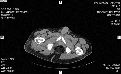
Fig. 8.2


Soft-tissue damage to the gluteal region. It is not clear that the wound extends to the anorectum. Clinical and endoscopic examination is required
Related posts:


Stay updated, free articles. Join our Telegram channel
Full access? Get Clinical Tree
