Pediatric Human Immunodeficiency Type 1 Infection
William V. Raszka JR., MD, FAAP
INTRODUCTION
Reports of unusual infections, such as Pneumocystis carinii pneumonia (PCP) and mucosal candidiasis in previously healthy homosexual men were first published in 1981 (Gottlieb, Schroff, Schanker, et al., 1981). Why patients developed such profound immunodeficiency was not known until 1983 when French and American researchers working independently announced that a retrovirus, now known as the human immunodeficiency virus (HIV), was the etiologic agent that led to the acquired immunodeficiency syndrome (AIDS) (Gallo, Salchuddin, Popovic, et al., 1984; Laurence, Brun-Vezinet, Scuter, et al., 1984). Once primarily a disease of homosexual men, HIV has spread throughout all segments of the human population. In some parts of sub-Saharan Africa, up to 40% of women of childbearing age are HIV infected. The economic and human losses are tremendous. Societies are losing important wage earners, infant mortality rates are rising in the most severely affected areas, and health care systems are strained or overwhelmed. Perhaps HIV will alter human evolution as no disease has since bubonic plague.
Knowledge about and perception of HIV infection have changed dramatically in the past decade. Dismal pessimism, followed by optimism that HIV infection could be “cured,” has given way to the notion that with adequate resources, HIV infection can be controlled although not eradicated. New prevention strategies and innovative treatment programs have decreased perinatal transmission rates from 25% to 8% (Connor, Sperling, Gelber, et al., 1994). Powerful combination drug regimens for the treatment of HIV infection have improved AIDS-free survival times in both pediatric and adult patients. Changes in the guidelines for drug development and approval that mandate testing in children have led to expanded access to new and more powerful antiretroviral drug regimens.
Although understanding and management of HIV have improved, enormous challenges remain. Behaviors that lead to an increased risk of HIV infection, such as sexual activity at an early age, multiple sexual partners, and drug use, are notoriously difficult to change. Ensuring that all HIV-infected women of childbearing age are identified and treated appropriately is difficult in the United States and beyond the abilities of most health care infrastructures in developing nations.
PATHOLOGY
HIV, or more appropriately, HIV-1, is a member of the Lentivirus genus in the family Retroviridae. Retroviruses contain two copies of single-stranded RNA genome. A unique feature of all retroviruses is their use of reverse transcriptase, an RNA-dependent DNA polymerase, to replicate genetic material. Other primate lentiviruses, such as simian immunodeficiency virus and its subspecies, are similar in structure to HIV but have important genetic and functional differences.
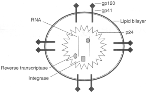
The mature HIV virion consists of three layers: an outer lipid bilayer studded with surface (GP120) and transmembrane (GP41) glycoprotein complexes; a middle layer consisting of matrix, internal capsular, and nuclear capsid proteins; and an internal region, which contains two copies of single-stranded RNA, multiple copies of viral-encoded reverse transcriptase, integrase, and RNAse H. Figure 65-1 illustrates the mature HIV virion.
The HIV-1 genome is organized similarly to that of other retroviruses but is more complex. The 9-kB RNA virus is flanked by long terminal repeat sections, which serve as both promoter and binding sites for host and viral transactivating factors. The genome contains structural (gag and env) and viral enzyme (pol) genes that encode precursor proteins that produce multiple gene products after intracellular processing. The env gene encodes surface glycoproteins GP120 and GP41 that are critical for binding to the CD4+ cell receptor. The gag region encodes nuclear capsid core and matrix proteins, while the pol gene encodes reverse transcriptase, protease, and integrase enzymes. The HIV-1 genome also has at least six other regulatory genes not found in nonprimate retroviruses whose products play critical roles in controlling viral expression, trafficking of viral gene products within the affected cell, and viral infectivity.
The primary binding site for the HIV virion is the CD4+ receptor. Cells that express CD4+ receptors include CD4+ T-cell lymphocytes and cells of the monocyte or macrophage line. While the CD4+ receptor site is a critical binding site, chemokines that function as coreceptors have been identified. All HIV-1 strains use CCR5 or CXCR4, members of the chemokine receptor family, as coreceptors. Lymphocytes from people who do not express CCR5 are relatively resistant to infection, rendering certain population groups inherently more susceptible or resistant to HIV viral infection and replication (Liu et al., 1996; Huang et al., 1996). Globally, the allelelic frequency of CCR5 deletion ranges from 0.5% in African and Asian populations to 20.9% in Ashkanazi Jewish populations (Martinson, 1997).
After binding to the CD4+ receptor complex and other receptors, the HIV virion uncoats and the HIV genome and regulatory enzymes enter the host cell. In the cytoplasm of the host cell, viral-encoded reverse transcriptase transcribes the single-stranded HIV RNA into complementary DNA. Multiple error mechanisms in reverse transcription lead to frequent mutations and significant HIV genomic diversity. In adults with established HIV disease, approximately 1010 new CD4+ T-cells are infected each day, making it possible that a mutation will occur at each point along the HIV-1 genome between 104 and 105 times per day (Perelson, Neumann, Markowitz, Leonard, & Ho, 1996). One consequence
of this genomic diversity is the production of antiretroviral-resistant strains.
of this genomic diversity is the production of antiretroviral-resistant strains.
Once proviral HIV DNA has been synthesized, it is integrated into the host DNA through the actions of viral-encoded integrase. Integrated proviral HIV DNA cannot be eradicated and represents a potential long-lived reservoir for viral persistence. Once integrated, several host transcriptional and viral activation factors stimulate viral replication or transcription. Newly produced proteins and genomic material aggregate near the cytoplasmic side of the cell membrane. One of the last enzymatic steps before budding from the cell membrane involves cleavage of terminal RNA sequences by a viral-encoded protease. Failure to complete this step leads to the production of nonreplicative virions.
Transmission
HIV may be transmitted either as a free virion or as integrated provirus within donated cells. Children become infected with HIV by exposure to infectious human, usually maternal, body fluids. To date in the United States, more than 90% of all pediatric patients with AIDS have acquired their infection perinatally (Centers for Disease Control and Prevention [CDC], 1998b). In 1997, only 9 of 473 pediatric patients diagnosed with AIDS had hemophilia, a coagulation disorder, or had received a blood transfusion, blood component, or tissue. Almost all new cases of HIV infection in children younger than 13 years will be the result of perinatal transmission.
• Clinical Pearl
HIV transmission associated with casual household contact or closed-mouth kissing is negligible (Courville et al., 1998). Open-mouth (“French”) kissing, particularly if the gingiva are inflamed or bloody, very rarely has been associated with transmission.
Biting very uncommonly results in transmission and only does so after extensive tearing and bleeding.
Contact with nonbloody saliva, urine, feces, tears, sweat, or biting insects has not resulted in HIV transmission (CDC, 1998d).
Perinatal Transmission
The pathogenesis of perinatally acquired HIV-I infection differs from that of adult infection in several significant ways. The developing immune system of the infant is not as effective at controlling viral replication. After infection, the number of HIV-1 RNA copies increases dramatically to 105 to 107 copies/mL (Palumbo et al., 1995; Shearer et al., 1997). Plasma HIV RNA levels decrease very gradually over the first 1 to 2 years of life, but mean plasma HIV RNA levels remain much higher than in adults. The reason for the persistently high viral levels in children is not known. Possible mechanisms include a larger pool of permissive host cells, relatively high thymic mass relative to body size, and an immature host immune response (Wilfert et al., 1994). Similar to adults, in children older than 2 years, plasma viral load at steady state is inversely related to long-term prognosis (Palumbo et al., 1998).
Perinatal transmission, often called vertical transmission, can occur at any time during pregnancy, at the time of delivery, or after birth.
Failure to detect HIV in infants born to HIV-infected women in the first weeks of life, with subsequent detection of virus in these infants at 1 to 3 months of age, demonstrates that intrapartum transmission occurs (Rogers et al., 1989; Dunn et al., 1995). The overall risk of HIV transmission from an untreated HIV-infected mother to her infant is approximately 25%, with published ranges from 13% to 39% (Connor et al., 1994; European Collaborative Study [ECS], 1992b; Tovo, deMartino, & Gabiano, 1992).
• Clinical Pearl
Appropriate antiretroviral therapy in the mother and the infant can decrease the risk of perinatal transmission by approximately two thirds.
Risk factors associated with an increased rate of perinatal transmission include a high maternal plasma copy number of HIV RNA (viral load); low maternal CD4+ lymphocyte cell count or percentage; advanced maternal HIV disease; maternal coinfection, such as chorioamnionitis or syphilis; premature delivery; second born of a twin delivery; prolonged rupture of membranes; and vaginal delivery. Although viral load best correlates with the risk of perinatal transmission, no single factor is predictive of HIV transmission.
Infectious HIV virions have been isolated from breast milk obtained from HIV-infected women. Studies in African populations have demonstrated that up to 30% of perinatal HIV transmission can occur postpartum from breast-feeding (Datta et al., 1994; Leroy et al., 1998). Numerous reports have documented HIV transmission through breast milk to infants born to HIV-negative mothers.
• Clinical Pearl
Because of the potential impact of breast-feeding on the overall mother-to-child transmission rate, bottle-feeding is currently recommended for all infants born to HIV-infected mothers in the United States and other industrialized countries (American Academy of Pediatrics [AAP], Committee on Pediatric AIDS, 1998).
In nonindustrialized countries, the issue is more controversial. Currently, the World Health Organization (WHO) recommends that children born to HIV-infected mothers in less developed parts of the world breast-feed, because perinatal morbidity and mortality rates are higher in children who are not breast-fed (HIV and Infant Feeding, 1999).
Sexual Abuse as a Means of Transmission
Sexual abuse is a rare mode of HIV transmission. Of 9136 children reported with HIV or AIDS through December 1996, only 26 had been sexually abused. Of these, 17 had confirmed and 9 suspected exposure to HIV (Lindegren et al., 1998).
Pathogenesis
Variation in the biologic properties of HIV isolates, such as cytotropism, syncytium-inducing capacity, and replication rate, may influence pathogenecity (DeRossi et al., 1993). Cells with integrated HIV DNA may produce huge numbers of HIV virions. The initial viremia leads to seeding of the virus throughout the body. HIV infects lymphoid organs very early in the disease. Follicular dendritic cells trap immune complexes of virus, antibody, and complement in the germinal centers of lymph nodes. This microenvironment is highly conducive to ongoing HIV infection of and replication in CD4+ lymphocytes as they migrate through lymphoid tissue, come into close contact with the follicular dendritic cells, and are activated (Health, 1995). Free virus and latently infected cells may remain sequestered within the lymphoid tissue and serve as a reservoir for persistent viral infection. Host immune responses, particularly the development of HIV-specific cytotoxic T cells, play a critical role in controlling viral replication (Musey et al., 1997). HIV-specific antibodies are detected only after the viremia has declined, suggesting that antibodies are not as critical to controlling viral replication. Why or how high-level viral replication continues despite HIV-specific cytotoxic T-cell lymphocyte production is unclear.
After widespread dissemination and high-grade viremia, the host immune response is effective at reducing the level of viremia. Approximately 6 months after infection in adults, viral load measured by plasma RNA polymerase chain reaction (PCR) reaches a steady state (Pantaleo, Graziosi, Fauci, 1993). This steady state stage may last 10 to 12 years and is often termed the time of clinical latency. This concept of immunologic latency is inaccurate. Although the patient experiences few clinical symptoms, rapid CD4+ cell turnover is occurring with high viral replication, and the patient experiences a steady incremental decline in CD4+ lymphocyte cell counts. The half-life of HIV-infected CD4+ cells may only be 2 days, and an estimated 1.8 × 10⁹ cells are destroyed daily (Ho et al., 1995). Maintaining CD4+ lymphocyte cell counts eventually becomes impossible in the face of continued or increased viral replication. The patient’s ultimate prognosis depends on how effective the host immune response has been in controlling viral replication. In adults, patients whose host immune response leads to a low level of viral replication and low plasma HIV viral load have a longer life expectancy than those who are unable to control viral replication as well (Mellors et al., 1995). This effect is independent of the CD4+ lymphocyte count.
The exact mechanism by which HIV replication leads to CD4+ lymphocyte cell death is not known. Possible mechanisms include direct lysis; induction of programmed cell death (apoptosis); host-specific immune responses, such as antibody-dependent cellular toxicity mediated by natural killer cells; and disruption of normal immunoregulatory pathways (Ameisen, Estaquier, Idziorek, & De Bels, 1995). Disease progression is associated with degeneration of the germinal center follicle cell network and disruption of lymphoid architecture (Cohen et al., 1995). Destruction of the lymphoid tissue impairs the host’s ability to control viral replication effectively, thereby allowing increased viral replication and further destruction of lymphoid tissue. With progressive lymphoid tissue destruction, the host’s ability to replace CD4+ lymphocytes is further compromised.
EPIDEMIOLOGY
Understanding the epidemiology of HIV infection is sometimes confusing. Until recently, only AIDS (which is at one end of a continuum of clinical syndromes associated with HIV infection) was a reportable condition. As of December 1997, only 29 states conducted surveillance for HIV infection among children. The CDC has published two separate classification systems for pediatric HIV infection. The 1987 AIDS case definition is used for purposes of surveillance and reporting (CDC, 1987). The CDC has developed a separate classification system to describe the spectrum of HIV disease in pediatric patients (CDC, 1994). Patients are classified into mutually exclusive categories according to three parameters: (1) infection status, (2) clinical status, and (3) immunologic status. CD4+ lymphocyte counts and percentage are used to gauge the degree of immunosuppression. Table 65-1 presents immunologic categories of pediatric HIV infection.
Once classified, an HIV-infected infant or child may not be reclassified in a less severe category even if antiretroviral therapy leads to clinical or immunologic improvement. The expanded definition for AIDS in adolescents and adults, which became effective in 1993, does not apply to individuals younger than 13 years of age (CDC, 1992). AIDS case definitions for children and adults are similar except that lymphoid interstitial pneumonitis and multiple or recurrent serious bacterial infections are AIDS-defining conditions only for children.
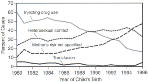
The epidemiology of pediatric HIV infection is greatly influenced by the course of the epidemic in childbearing women because the major route of transmission of HIV to children is through perinatal exposure. In most parts of the world, including North America, women of childbearing age (15–44 years) represent one of the fastest growing groups with AIDS. In the United States, women counted for 20% of adult AIDS cases reported in 1996, compared with less than 10% in the 1980s (CDC, 1996). While AIDS case rates have risen in women of all ethnic backgrounds, African American and Hispanic women have higher AIDS case rates than Caucasian women (Whortley & Fleming, 1997). Women with heterosexual contact as the only risk factor for HIV increased
from 14% in 1982 to 35% in 1993. Heterosexual contact is now the primary means by which women acquire their infection. Figure 65-2 presents information about perinatally acquired AIDS in the United States between 1980 and 1996.
from 14% in 1982 to 35% in 1993. Heterosexual contact is now the primary means by which women acquire their infection. Figure 65-2 presents information about perinatally acquired AIDS in the United States between 1980 and 1996.
 Figure 65-2 Mother’s exposure category by year of child’s birth for perinatally acquired AIDS, 1980–1996, in the United States. Adapted from Centers for Disease Control and Prevention. (1997). HIV/AIDS Surveillance Report 9, 1–39. |
Pediatric HIV/AIDS has become a leading cause of death in children worldwide. By June 1998, an estimated 2.7 million children worldwide had acquired HIV infection. Of these, more than 1.7 million had progressed to AIDS, and 1.4 million had died (UNAIDS, 1998). Worldwide, more than 1000 children are newly infected with HIV each day. U.S. Bureau of the Census statistics suggest that in many areas, HIV infection has led to increased infant mortality rates and that this effect is expected to increase over the coming decade (Stanecki & Way, 1997). However, significant regional variations exist. Sub-Saharan Africa accounts for the greatest proportion of cases. Although the epidemic began later in Asia and India, the epidemic in this region may overshadow all others because of its large population. HIV infection is well established in Latin America and the Caribbean. Published HIV rates in the Middle East have remained low.
AIDS is the seventh leading cause of childhood mortality in the United States in the age group 1 to 4 years, the fourth leading cause of death among African American children ages 1 to 4 years, and the fifth leading cause of death among Hispanic children. In some areas, such as New York, New Jersey, and Florida, HIV has become one of the top three leading causes of childhood mortality (National Institute of Allergy and Infectious Diseases, 1996). As of September 30, 1997, perinatal transmission of HIV accounted for 7310 (1%) of the 626,334 total AIDS cases in adults and children reported to the CDC by state and territorial health departments (CDC, 1998b). Perinatally acquired pediatric AIDS cases have been reported from 48 states, the District of Columbia, Puerto Rico, and the Virgin Islands. Five states or territories account for 64% of all perinatally acquired AIDS cases: New York (27%), Florida (17%), New Jersey (9%), California (6%), and Puerto Rico (5%). The vast majority of cases (85%) were diagnosed in metropolitan areas with populations of more than 500,000 people. Ethnic minorities are overrepresented, because only 14% of cases were in non-Hispanic Caucasian patients (CDC, 1997b). Table 65-2 illustrates these data.
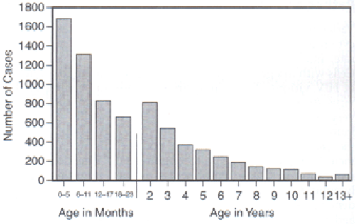
The median age at the time of AIDS diagnosis in children is 17 months. Forty percent of cases are diagnosed in children younger than 1 year, 47% in children ages 1 to 5 years, and 13% in children age 6 years and up. Figure 65-3 demonstrates data for diagnosis of pediatric AIDS by age in the United States between 1982 and 1997.
 Figure 65-3 Perinatally acquired AIDS Cases by Age at Diagnosis, 1982–1997, in the United States. Adapted from Centers for Disease Control and Prevention. (1997). HIV/AIDS Surveillance Report 9, 1–43. |
Contrary to what is happening in many other parts of the world, the number of U.S. children with newly diagnosed AIDS or new perinatally acquired HIV infection is declining. Public health efforts to identify all HIV-infected pregnant women and implementation of zidovudine-based antiretroviral treatment regimens during pregnancy and in the offspring of HIV-infected mothers have led to a substantial decline in the reported incidence of pediatric AIDS cases in the past several years (Simonds et al., 1998). From 1984 through 1992, the estimated number of children with perinatally acquired AIDS diagnosed each year increased, then declined 43% from 1992 to 1996. During this time, declines were similar by race or ethnicity, for different regions of the United States, and in urban and rural areas. The declines were largest among children in whom AIDS was diagnosed at younger ages (CDC, 1998b).
HISTORY AND PHYSICAL EXAMINATION
Infants and children infected with HIV may develop many disparate clinical problems. Some may be directly attributable to HIV infection. Others, such as most infectious disease complications, are the result of HIV-induced immunosuppression. Before widespread maternal HIV testing and antiretroviral use, approximately 50% of children with vertically acquired HIV infection were symptomatic by 8 months and 80% by 2 years of age (Scott et al., 1989). Prospective and retrospective data suggest that two populations of children exist. Approximately 25% of children have rapid progression and death within the first few years, while the remainder survive early childhood and have a clinical course more consistent with adult infection. These children have a mean survival time of more than 9 years (Barnhart et al., 1996).
In adolescents and young adults, the primary viremia associated with HIV infection presents as a mononucleosis-like illness. This illness, called the acute retroviral syndrome, is characterized by fever, fatigue, myalgia or arthralgia, adenopathy, pharyngitis, diarrhea, headache, and rash, and it lasts 1 to 2 weeks (Quinn, 1997) Congenital HIV infection is not associated with any distinguishing clinical or morphologic features (Connor et al., 1994). The acute retroviral syndrome is not as well described in infants. Infants may present only with fever or irritability only.
DIAGNOSTIC STUDIES
Lymphoid interstitial pneumonitis/pulmonary lymphoid hyperplasia (LIP/PLH) is a chronic pulmonary disease of unknown etiology characterized by diffuse peribronchiolar lymphoid nodules. LIP/PLH is second only to PCP among pediatric AIDS-defining conditions. Approximately 20% of children present with LIP/PLH as their AIDS indicator disease. Although the incidence has declined slightly, LIP/PLH has remained surprisingly common, even in the era of highly active antiretroviral therapy. The pathogenesis of LIP/PLH is not known. Initially, patients are generally asymptomatic but may have generalized lymphadenopathy. Characteristic clinical features of late disease include tachypnea, cough, wheezing, hypoxemia, and digital clubbing. The most common radiographic findings include a diffuse reticulonodular infiltrate with or without hilar adenopathy. A presumptive diagnosis of LIP can be made based on typical radiographic findings persisting for 2 months that are unresponsive to antimicrobial therapy or, more definitively, by lung biopsy. Although features of LIP/PLH and PCP can be similar, patients with LIP/PLH usually are older, have a more insidious disease onset, and do not have fever or crackles on auscultation.
Diagnosing HIV infection in infants is more complicated than identifying HIV infection in an adult. Immunologic, virologic, and clinical criteria have been published (CDC, 1994). Because maternally derived anti-HIV IgG antibodies may persist for as long as 18 months after delivery, IgG-based serologic assays do not reliably confirm HIV infection in infants less than 18 months old.
• Clinical Pearl
Two or more negative anti-HIV IgG antibody test results obtained at greater than 6 months of age, with an interval of at least 1 month between tests, excludes HIV infection in any child of any age without clinical evidence of HIV infection, unless the child is hypogammaglobulinemic or newly infected before the development of an immunologic response.
HIV-specific IgM antibody tests lack sensitivity and specificity and are not available. HIV-specific IgA antibodies have been used to diagnose HIV infection in infants at least 3 months old (Kline et al., 1994). Although used infrequently in the United States, IgA testing is inexpensive and easy to perform, making this a useful tool in many parts of the world. Display 65-1 presents guidelines for making a laboratory diagnosis of HIV in infants and children.
DISPLAY 65–1 • Laboratory Diagnosis of HIV Infection in Infants and Children
Laboratory Criteria for Considering a Child HIV Infected
Infants younger than 18 months:
Two separate HIV tests by culture, polymerase chain reaction, or p24 antigen* are positive.or
The child is HIV antibody-positive and meets criteria for AIDS diagnosis based on the 1987 AIDS surveillance case definitions (CDC, 1987).
Children ages 18 months and older:
Repeated HIV-antibody tests by reactive enzyme immunoassay and confirmatory tests (ie, Western blot or immunofluorescence assay) are positive,
or
The child meets the criteria in 1A.
Laboratory Criteria for Considering a Child HIV Negative
An infant or child is HIV negative (a seroreverter if born to an HIV-infected mother) if the child has the following:
Two or more HIV antibody tests performed between 6 and 18 months of age or one HIV antibody test after 18 months of age is negative,
or
Two separate polymerase chain reaction tests* are negative,
and
Child has not had an AIDS-defining condition.
Footnotes
HIV = human immunodeficiency virus; AIDS = acquired immunodeficiency syndrome.
*Both determinations were performed at or beyond 1 month of age and at least one at or beyond 4 months of age.
Adapted from Centers for Disease Control and Prevention. (1994). 1994 Revised Classification System for human immunodeficiency virus infection in children less than 13 years of age. Morbidity and Mortatlity Weekly Report, 43(RR-12), 1–10.
In the United States, nonserologic tests are used for the laboratory diagnosis of HIV infection in infants younger than 18 months. Both HIV culture and PCR are sensitive and specific assays for HIV infection. HIV culture, however, is expensive and time-consuming and is not often used. PCR, which amplifies either HIV RNA from the plasma or proviral DNA from infected mononuclear cells, is currently the diagnostic test of choice. With PCR, HIV infection can be diagnosed definitively in most infants by age 1 month and in virtually all infants by 6 months. A meta-analysis of published data from 271 HIV-infected children showed that 38% had positive HIV DNA PCR test results by 48 hours of age and 93% by the end of the second week (Dunn et al., 1995). In a review of 1209 samples taken at 1 to 36 months of age from 483 HIV-exposed infants, a single PCR assay was 95% sensitive and 97% specific in diagnosing HIV infection (Kline et al., 1994).
• Clinical Pearl
To date, maternal or infant zidovudine therapy has not delayed the detection of HIV infection in infants and does not decrease the sensitivity or predictive values of most virologic assays (Conner et al., 1994; Kovacs et al., 1995).
Although the CDC recommends the use of DNA-based PCR tests to diagnose HIV infection, recent data would suggest that RNA-based PCR testing modalities, which are ubiquitous, may be equally sensitive (Steketee et al., 1997). Standard and immune complex dissociated p24 antigen tests are highly specific for HIV infection but are not sensitive compared with other virologic assays and are associated with an unacceptably high false-positive rate in infants younger than 1 month (Nesheim et al., 1997).
Stay updated, free articles. Join our Telegram channel

Full access? Get Clinical Tree


