- •
Anterior chest wall blocks are used as alternatives to thoracic epidurals and paravertebral blocks for breast surgeries and procedures involving the anterior chest walls.
- •
Serratus plane blocks and supraclavicular blocks may be required for more extensive surgeries.
- •
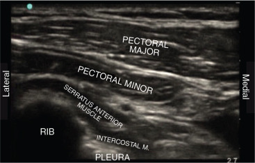
Pectoralis major, pectoralis minor, and serratus anterior muscles are the main landmarks for the PEC blocks at the level of the fourth and fifth ribs.
- •
Bilateral pecto-intercostal blocks are required for sternal wound incisions.
- •
All fascial plane blocks need ultrasound guidance, skill with ultrasound-guided procedures, and larger volume of local anesthetics.
Perspective and background
The introduction of ultrasound to regional anesthesia allowed identification of various interfascial planes between the different muscles, including the chest wall musculature. Some of the interfascial planes (between the investing fascias of the muscles) are considered neurovascular potential spaces, hosting the different nerves and vessels supplying the chest wall. Along the course of the intercostal nerves, they branch and communicate with the adjacent intercostal nerves as well as other pectoral nerves when crossing between those different interfascial planes.
The ventral rami of the thoracic spinal nerves (T2–T9) pierce the intercostal muscles giving lateral and anterior cutaneous branches that further divide into posterior, anterior, medial and lateral branches, providing sensory innervations to the anterior and lateral aspects of the chest wall including the breast (T4–5–6) . The apex of the axilla is innervated by the intercostobrachial nerve, a branch of T2 spinal nerve. These multiple divisions of the spinal nerves communicate with the branches of the adjacent spinal nerves through the neurovascular planes existing between the muscles of the chest wall, mainly pectoralis major, pectoralis minor, and serratus anterior muscles, bounded by the fascial layers covering the muscles (interfascial planes). Those planes also host branches of the brachial plexus along their course in the chest; mainly medial pectoral nerve (C8–T1), lateral pectoral nerve (C5–7), long thoracic nerve (C5–7), and thoracodorsal nerve (C6–8) ( Figs. 30.1–30.4 ).