Fig. 5.1
Critical view of safety as defined by strassberg
There is no additional preparation needed than that done for a laparoscopic or open cholecystectomy.
Consent for a laparoscopic cholecystectomy should always include possible conversion to an open procedure, bile duct/vascular injury, and need for a drain.
Operative Strategy
Partial cholecystectomy can be performed using either an open or laparoscopic technique. We will address the open technique but the general principles can be applied to a laparoscopic approach. PC allows for amputation of either just the anterior wall of the gallbladder or removal of the gallbladder at the level of the infindibulum. Once resected the contents of the gallbladder and cystic duct are cleared, the mucosa obliterated and the stump closed.
Operative Technique
Make a Kocher incision two finger breaths below the right costal margin, incising fascia and rectus abdominis and lateral muscles. Maintain hemostasis of abdominal wall vasculature as the peritoneal cavity is entered.
One may need to locate and divide the falciform ligament to aid exposure and assist with retraction.
A self-retaining retractor should be used to facilitate visualization and allow the surgeon and assistant use of both hands. The authors prefer a Thompson or Omni retractor; however, a Bookwalter may be used.
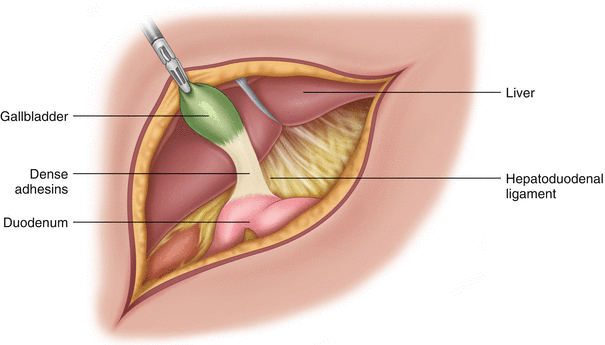
Any adhesions not already incised should be taken down allowing clearance of the infindubulum as low as safely possible (Fig. 5.2).


Fig. 5.2
Dense adhesions making a traditional cholecystectomy high risk for biliary injuries
Gallbladder distension may be relieved with either needle or suction decompression. Bile leakage should be minimized by occluding the opening with a stitch or hemostat.
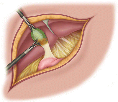
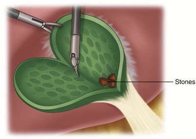
If dissection is difficult between the gallbladder and liver bed, it is possible to leave the posterior wall of the gallbladder intact. The gallbladder is first entered approximately 1 cm from liver edge and the full thickness of the gallbladder is transected parallel to the liver to a safe level (Figs. 5.3 and 5.4).


Fig. 5.3
Entering the gallbladder at the fundus. One can appreciate the lack of a dissection plane between the liver and the gallbladder

Fig. 5.4
Transection is continued through the full thickness of the gallbladder wall parallel to the liver surface. Stones can sometimes be appreciated
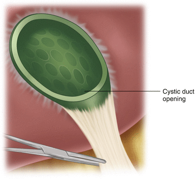
The transection is then brought around the anterior surface of the gallbladder exposing both the posterior wall and infindibulum mucosa (Fig. 5.5).


