Fig. 16.1
This CT scan illustrates a gastric volvulus of a paraesophageal hernia. The “swirl” is seen within the stomach in the chest. © Dale Dangleben, MD
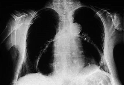
Fig. 16.2
Large paraesophageal hernia is seen here on chest X-ray overlying the cardiac silhouette. © Dale Dangleben, MD
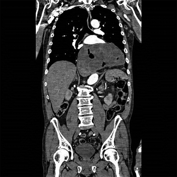
Fig. 16.3
Large paraesophageal hernia is seen here on CT scan. Most of the stomach is within the thoracic cavity. © Dale Dangleben, MD
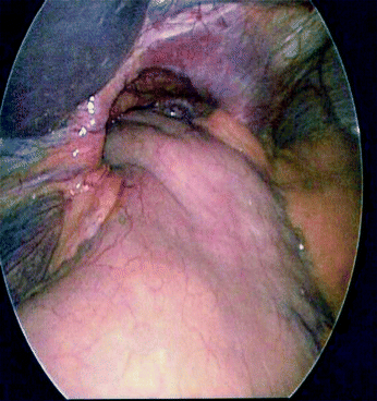
Fig. 16.4
Laparoscopic view of a large paraesophageal hernia. © Dale Dangleben, MD
References
1.
Bhayani NH, Kurian AA, Sharata AM, Reavis KM, Dunst CM, Swanstrom LL. Wait only to resuscitate: early surgery for acutely presenting paraesophageal hernias yields better outcomes. Surg Endosc. 2013 Jan;27(1):267–71.
Related posts:


Stay updated, free articles. Join our Telegram channel

Full access? Get Clinical Tree


