Figure 38.1
Initial dissection for pancreaticoduodenectomy. A curvilinear, subcostal incision is first carried out over the right upper quadrant and is subsequently extended across the midline as far as is necessary for adequate surgical exposure. Excessive care must be taken to maintain meticulous hemostasis, especially in the jaundiced patient. After making the skin incision, the rectus muscles are carefully transected with bipolar electrocautery (shown above)
Before irreversible steps are taken, a thorough exploration of the abdomen must be carried out to determine the location and extent of the pathologic process and to detect any evidence of tumor spread outside the limits of resection. The liver and all serosal surfaces should be carefully examined for metastatic spread or peritoneal dissemination. In addition, metastatic spread to the periportal and celiac axis lymph nodes, the root of the transverse mesocolon, the region above the pancreas, and the hepatoduodenal ligament should be sought by careful examination. The discovery of involved peripancreatic lymph nodes, even along the cephalad border of the pancreas near the portal vein, does not preclude resection, although it worsens prognosis [9]. Ultrasound may be helpful in ruling out metastatic spread to the liver.
Once disseminated disease has been ruled out, the surgeon proceeds with mobilization of the duodenum and head of the pancreas by the Kocher maneuver. Dissection of the lateral peritoneal attachments of the duodenum, which facilitates inspection of the duodenum, head of the pancreas, and periampullary tumor is usually bloodless; an avascular cleavage plane can be easily developed as the posterior wall of the pancreas is bluntly separated from the underlying vena cava and right kidney. Extensive kocherization should be performed to allow the surgeon to be comfortable that there is no extension of tumor beyond the uncinate process. Special care should be taken to identify and preserve the right gonadal vein, which often runs parallel to the inferior vena cava at this point in the retroperitoneal dissection Fig. 38.2. Further mobilization of the second and third portion of the duodenum is carried out to adequately determine resectability of the lesion.

Figure 38.2
Relationship of retroperitoneal structures encountered in the early stages of pancreaticoduodenectomy (Picture taken from patients left side). Once disseminated disease has been ruled out, the surgeon may proceed with mobilization of the duodenum and head of the pancreas by the Kocher maneuver. With adequate duodenal mobilization, the surgeon should identify and preserve the right gonadal vein, which arises from and often runs parallel to the inferior vena cava at this point in the retroperitoneal dissection
Before concluding that the lesion is resectable, the lesser sac must be entered to facilitate visualization and mobilization of the pancreas. The greater omentum is retracted upward and the gastrocolic ligament is incised all the way to the splenic flexure, allowing entry into the lesser sac. The right gastroepiploic artery and vein are identified and a thorough evaluation of potential metastases above the pancreas and adjacent to the celiac axis lymph nodes should be performed. The middle colic vein with its origin at the superior mesenteric vein should be identified and confirmed to be free of tumor involvement. The peritoneal attachments at the inferior border of the pancreas are incised and a cleavage plane over the superior mesenteric vein and behind the pancreas (the so-called “tunnel of love”) is developed Fig. 38.3. Development of the subpancreatic tunnel will permit the surgeon to continue dissecting behind the pancreas and over the portal vein, to be certain it is not involved with tumor.
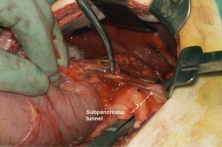
Figure 38.3
Development of the subpancreatic tunnel. The peritoneal attachments at the inferior border of the pancreas are incised and a cleavage plane over the superior mesenteric vein and behind the pancreas (the so-called “tunnel of love”) is developed. Development of the subpancreatic tunnel allows the surgeon to dissect posterior to the pancreatic neck and separate the tissues from the underlying portal vein. It is vital that the portal vein be identified at this portion of the case to be certain it is not involved with tumor. Note the clamp is behind the pancreas, over the SMV/PV
After the tumor is deemed resectable, irreversible steps can be taken. The gallbladder should be removed to prevent late complications from gallstone formation. Using electrocautery, the gallbladder is carefully dissected from the hepatic fossa. Meticulous hemostasis, especially in the jaundiced patient, should be maintained within the liver bed. The cystic artery is identified, doubly clipped, and transected. Dissection should continue to the common bile duct where it is encircled with a vessel loop for subsequent transection (Fig. 38.4). The surgeon then proceeds to ligate the blood supply necessary for antrectomy. The right gastric artery is identified, ligated with 2-0 silk sutures, and subsequently transected. Next, the gastroduodenal artery (GDA), passing inferiorly from the hepatic artery at the point where the portal vein passes posterior to the pancreas, should be suture ligated with 4-0 Prolene sutures. Just before ligating and dividing the GDA, the vessel should be occluded with a vessel loop or bulldog clamp to ensure adequacy of the hepatic artery pulse. At this point, important anatomic anomalies such as a replaced right hepatic artery originating from the superior mesenteric artery (SMA) or a replaced common hepatic artery off the SMA, should be evaluated by palpating for an arterial pulse behind the common bile duct through the foramen of Winslow. Following this, the right gastroepiploic vessels are ligated and tied.
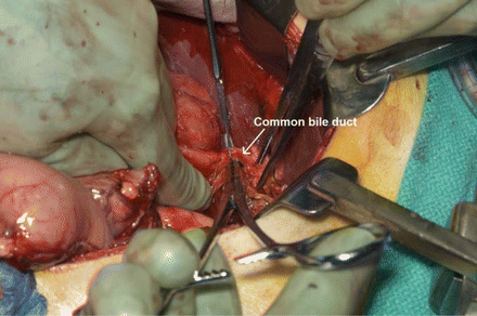
Figure 38.4
Identification and division of the common bile duct. The gallbladder should be removed to prevent late complications from gallstone formation. Using electrocautery, the gallbladder is carefully dissected from the hepatic fossa. Meticulous hemostasis, especially in the jaundiced patient, should be maintained within the liver bed. The cystic artery is identified, doubly clipped, and transected. Dissection should continue down the common bile duct, transecting it above the cystic duct
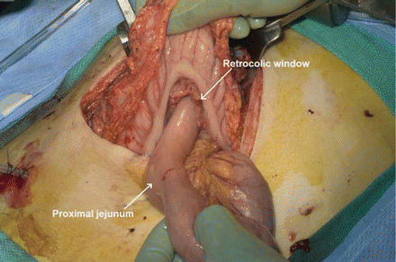
The removal of the antrum greatly assists in the subsequent exposure of the more difficult portion of the resection. After an area is cleared on both the greater and lesser curvature of the stomach, an antrectomy is performed using a GIA stapler. Once the stomach is transected, the remainder of the resection is carried out. The common hepatic duct is sharply transected just above the cystic duct. This not only allows the surgeon to perform a hepaticojejunostomy during the reconstructive phase of the procedure but also allows him or her to adequately visualize the portal vein. Attention is now directed toward mobilization of the upper jejunum. The transverse colon is flipped superiorly, allowing for adequate visualization of the jejunum and its mesentery. The upper jejunum may be grasped with Babcock forceps and the bowel held up in order to adequately visualize the vascular arcades supplying the jejunum. The ligament of Treitz, in its avascular plane, is taken down with cautery Fig. 38.5. Utilizing incisions made in the avascular portions of the mesentery, the jejunum is divided with a GIA stapler. The jejunal arcades are divided and ligated to facilitate mobilization of the upper jejunum. A small opening is made in the mesocolon underneath the SMV and the mobilized upper jejunum is passed through the retrocolic window.