Chapter 4 Other systems
Dental anaesthesia
The first dental GA was given by Cotton and Wells in 1844. Currently, 300 000 dental GAs are given per annum (70% children) and, until recently, numbers have been declining owing to less tooth decay.
Mortality is 1:150 000 (2 deaths/year), compared with 1:250 000 non-dental day-case GAs, and is usually due to respiratory difficulties or sudden cardiovascular collapse.
Recent deaths in the dental chair have prompted moves to stop dental anaesthesia being carried out in dental surgeries.
Department of Health 1990
This report discusses general anaesthesia, sedation and resuscitation in dental surgeries. Its main recommendations are:
Standards and Guidelines for General Anaesthesia for Dentistry
The Royal College of Anaesthetists 1999
Introduction
Background
Recommended standards
General anaesthesia should be limited to:
Patient assessment
The final decision as to the benefit:risk of a dental GA can only be made after consultation between the patient, anaesthetist and dentist.
Clinical setting
Risks of death are greater should a complication occur. There must be written protocols for the management of patients requiring resuscitation or transfer.
Equipment and drugs
Equipment should be appropriate to the dental setting and anaesthetic technique. Routine maintenance must be performed and checks of equipment must be made before use. Back-up equipment and resuscitation drugs must be available.
Techniques
General anaesthetic
Intubation. Avoid suxamethonium, which has high morbidity. High-dose alfentanil (30 μg/kg) together with propofol enables intubation without relaxant.
Indicated for mental handicap, prolonged or painful surgery and nasal airway obstruction. Use nasopharyngeal pack.
Cardiovascular side-effects
Dysrhythmias. These are common due to sympathetic and parasympathetic activity, high levels of endogenous catecholamines, halothane and airway obstruction with hypoxia and hypercarbia:
They mostly occur during surgery and are worse during trigeminal nerve stimulation. Atropine increases the incidence of dysrrhythmias.
Less common with i.v. induction than with gaseous induction. Less common with volatile agents other than halothane.
More common following recent Coxsackie B infection due to (?) viral myocarditis.
Arrhythmias may be the primary cause of high mortality seen with dental GAs.
Fainting. Only in conscious patients! May need large doses of atropine. Abandon procedure because CVS instability persists for up to 90 min.
High mortality in erect patients was attributed to unrecognized fainting, but mortality is the same in supine patients. Supine position is associated with more pharyngeal soiling. It is now recommended that all surgery should be performed in the supine position which gives better CVS stability.
Cartwright D.P. Death in the dental chair. Anaesthesia. 1999;54:105-107.
Department of Health. General anaesthesia, sedation and resuscitation in dentistry (Poswillo Report). London: HMSO, 1990.
Flynn P.J., Strunin L. General anaesthesia for dentistry. Anaesth Intens Care Med. 2005;6:263-265.
Royal College of Anaesthetists. Standards and guidelines for general anaesthesia and dentistry. London: RCA, 1999.
Worthington L.M., Flynn P.J., Strunin L. Death in the dental chair: an avoidable catastrophe? Br J Anaesth. 1998;80:131-132.
Anaesthesia for ear, nose and throat surgery
General anaesthetic problems
Ear surgery
Myringotomy
This is a short, relatively painless operation. Premedication is not usually necessary.
Chronic otitis media is often associated with upper respiratory tract infection. Morbidity not increased with uncomplicated URTI if mask rather than intubation used. Need postoperative O2 if oxygen saturation <93% on room air.
Middle ear surgery
Careful positioning is required to avoid obstruction of venous drainage of the head. Tympanoplasty and mastoidectomy usually require identification of facial nerve. Therefore, avoid long-acting neuromuscular blocking drugs.
Use hypotensive anaesthesia to minimize bleeding. Maximum recommended safe dose of adrenaline = 0.1 mg/10 min.
Avoid N2O which diffuses into the middle ear to cause expansion, or on cessation diffuses out of the middle ear to cause negative pressure and disruption of ossicles.
Nasal surgery
Anaesthesia of the nasal cavities
Moffatt’s solution
Give with head extended over trolley, with half into each nostril.
Sluder’s technique. Four applicators are dipped into adrenaline and cocaine solution, and placed on middle turbinates under anterior and posterior ends.
Packing of nasal cavities. Spray nasal cavities with 4–10% cocaine and then pack with gauze soaked in 4–5% cocaine solution.
Gauze must contact area behind middle meatus (greater and lesser palatine nerves) and ethmoidal plate (anterior ethmoidal nerve).
Throat surgery
Tonsillectomy
Avoid premedication if tonsils are large or there is a history of sleep apnoea. Use gas or i.v. induction. Either deep gaseous intubation (patient more drowsy postoperatively) or suxamethonium. Use throat pack and endotracheal tube. Spontaneous respiration tends to hypoventilation with risk of arrhythmias, especially with halothane.
Extubate awake (protective reflexes), with head-down in left lateral position.
Postoperative haemorrhage. Affects 0.5%; 75% of postoperative haemorrhages occur within 6 h of surgery. Main problems are:
Assessing the patient can be difficult. Tachycardia due to hypovolaemia may also be due to anxiety or pain. Blood loss is usually underestimated, as most is swallowed. Establish i.v. access, check BP sitting and lying (postural hypotension with hypovolaemia), check haematocrit and cross-match blood.
There are two approaches to induction:
Both approaches need a selection of laryngoscope blades, stylettes, range of ETTs, two suction units (one may become blocked with clot), emergency tracheostomy kit and tipping trolley.
Adenoidectomy
Usually performed in conjunction with tonsillectomy. Not as painful. May cause nasopharyngeal obstruction and obligate mouth breathing.
Obstructive sleep apnoea
Grossly hypertrophied tonsils can cause partial upper airway obstruction when awake and complete obstruction during sleep. Associated with obesity, micrognathia (e.g. Pierre–Robin syndrome) and neuromuscular disorders (e.g. cerebral palsy). May present as failure to thrive, snoring, daytime somnolence, developmental delay, recurrent chest infections and, if severe, cor pulmonale. Airway obstruction may persist post-tonsillectomy.
Peritonsillar abscess (quinsy)
The infected tonsil forms an abscess in the lateral pharyngeal wall with associated trismus and difficulty in swallowing. The abscess does not usually interfere with the airway, but there is a risk of rupture and aspiration of contents. Drainage under LA, otherwise treat as for epiglottitis. Consider tracheostomy under LA if abscess is likely to rupture on intubation.
National Patient Safety Agency
Reducing the Risk of Retained Throat Packs after Surgery, April 2009
This Safer Practice Notice applies to all members of theatre teams and aims to reduce the risk of throat packs being retained after surgery is completed. Throat packs are often inserted by anaesthetists or surgeons to:
However, if a throat pack is retained after surgery is completed, it can lead to obstruction of the patient’s airways. Data received by the Reporting and Learning System between 1 January 2006 and 31 December 2007 were analysed. A total of 38 incidents were identified, of which 24 were unintended retention of throat packs; one resulting in moderate harm.
Clinical risk managers responsible for anaesthesia and surgery should ensure that local policies and procedures are adapted to state that:
Anaesthesia and liver disease
Normal physiology
The liver receives 25% of cardiac output. Portal venous blood contributes 70% of total flow and 50–60% of oxygen. The portal venous system has little smooth muscle and is not as responsive to sympathetic tone as the hepatic artery. When portal flow decreases, hepatic artery flow increases. Autoregulation at a microvascular level is poorly developed in the liver but hepatocytes can extract more oxygen from the blood than can any other tissue.
Total hepatic blood flow is reduced by IPPV, PEEP, hypovolaemia, hypocarbia, general anaesthesia and epidurals.
Physiological changes in liver disease
CVS. Hyperdynamic circulation, ↓ systemic vascular resistance, portal hypertension, due to activation of renin–angiotensin system and intravascular and interstitial fluid accumulation. Alcoholic cardiomyopathy.
Respiratory. Restrictive lung disease, pleural effusions. Impaired hypoxic pulmonary vasoconstriction causing ↑ V/ scatter and ↑ shunting, together with impaired respiration from ascites, causing hypoxia.
Haematology. Anaemia, thrombocytopenia, coagulopathy.
Renal. Hepatorenal syndrome (especially with sepsis and ↑ bilirubin). Prerenal failure causes acute tubular necrosis.
GI. Oesophageal varices, delayed gastric emptying. Increased gastric volume and acidity.
CNS. Encephalopathy, cerebral oedema.
Metabolic. Metabolic and respiratory alkalosis, hyper- or hypoglycaemia. Hyponatraemia (usually dilutional).
Pharmacokinetic and pharmacodynamic changes
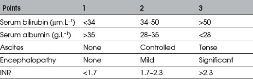
Assessment of surgical risk
Child’s (1963) classification assessed risk using albumin and bilirubin. Modified by Pugh et al (1973) (see Table 4.1). Perioperative mortality A<5%, B≈︀25%, C>50%.
Specific drugs
Anticholinergics. Little change in pharmacokinetics. Use normal doses.
Barbiturates. Increased sensitivity and prolonged excretion of thiopentone. Use <3–4 mg/kg.
Benzodiazepines. Lorazepam and oxazepam are metabolized by glucuronidation, which is minimally affected: therefore they are safe. Increased sensitivity to diazepam and midazolam due to impaired phase I reactions.
Propofol. Increased sensitivity. Use 2 mg/kg for induction.
Opioids. Increased sensitivity and prolonged half-life of morphine, diamorphine and pethidine. No change in remifentanil pharmacokinetics.
Muscle relaxants. Suxamethonium has prolonged action due to reduced plasma cholinesterase. Resistance to pancuronium due to increased VD of these drugs. Atracurium is probably the drug of choice due to spontaneous degradation and little change in pharmacokinetics. Action of vecuronium is prolonged even with mild liver disease.
Anticholinesterases. Normal doses of neostigmine may be used.
Local anaesthetics. Reduced metabolism of amides. Reduced plasma cholinesterase prolongs elimination of esters.
Inhalational agents. Halothane > enflurane decrease hepatic blood flow. Little effect with isoflurane/sevoflurane/desflurane. Avoid halothane because of risks of hepatitis. Sympathomimetic effects of N2O may reduce hepatic blood flow.
Other. Adrenaline and ephedrine increase hepatic blood flow. Sodium nitroprusside and β-blockers decrease hepatic blood flow.
Anaesthetic management
Preoperative
Assess cardiovascular and renal status. Optimize respiratory function with antibiotics, bronchodilators, physiotherapy and consider drainage of ascites. Correct coagulation with FFP, cryoprecipitate, vitamin K and platelets.
Maintenance
Use isoflurane/sevoflurane/desflurane in oxygen. High Fio2 needed because of pulmonary shunting. Avoid hyperventilation which increases mean intrathoracic pressure, thus reducing hepatic blood flow, accelerates formation of ammonia, causing hepatic encephalopathy, and increases urinary potassium loss through respiratory alkalosis. Hepatic blood flow is proportional to mean arterial pressure. Keep CVP <5 mmHg to reduce haemorrhage secondary to venous congestion.
Neuromuscular blockade with atracurium or pancuronium. Reversal with normal doses of anticholinesterases. Avoid rocuronium which has prolonged action.
Maintain high urine output to protect against renal failure. Avoid excess sodium load in i.v. fluids to prevent dilutional hyponatraemia.
Hepatic disease reduces synthesis of coagulation factors and inhibitors, and causes quantitative and qualitative platelet defects and hyperfibrinolysis. Massive haemorrhage a particular risk with cirrhosis, steatosis, and after chemotherapy. Exacerbated by acidosis, hypothermia and hypocalcaemia. Use of cell salvage in malignancy is controversial. Anti-fibrinolytics (e.g. tranexamic acid) reduces perioperative blood loss.
Postoperative
IPPV may be necessary until patient is rewarmed and stabilized. Remove lines as soon as possible to reduce risk of infection. Analgesia with small doses of opioids. Remifentanil avoids accumulation of active opioid metabolites. Regional block may reduce requirements, provided clotting is normal.
Watch for hypoglycaemia as a result of impaired hepatic mobilization of glucose. Secondary hyperaldosteronism is common (sodium and water retention and oedema), which is minimized by restriction of sodium intake and treated with diuretics.
Fabbroni D., Bellamy M. Anaesthesia for hepatic transplantation. Contin Edu Anaesth, Crit Care Pain. 2006;6:171-175.
Hartog A., Mills G. Anaesthesia for hepatic resection surgery. Contin Edu Anaesth, Crit Care Pain. 2009;9:1-5.
Lai W.K., Murphy N. Management of acute liver failure. Contin Edu Anaesth, Crit Care Pain. 2004;4:40-43.
Anaesthesia for ophthalmic surgery
Physiology
Aqueous humour
Made by ciliary plexus and secreted into anterior chamber. Absorbed through the trabecular meshwork into the canal of Schlem.
Intraocular pressure (IOP)
Normal value 15–25 mmHg. Once the eye is opened, IOP is equal to atmospheric pressure. Hypoxia, hypercapnia, coughing and vomiting all increase IOP.

Stay updated, free articles. Join our Telegram channel

Full access? Get Clinical Tree

