Figure 33.1
An infraumbilical, semicircular incision is made
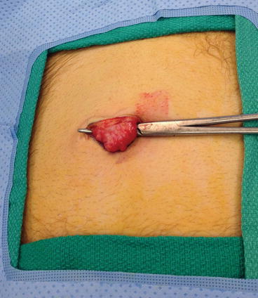
Figure 33.2
A clamp around the isolated umbilical stalk. Division of the stalk allows access to the hernia defect below
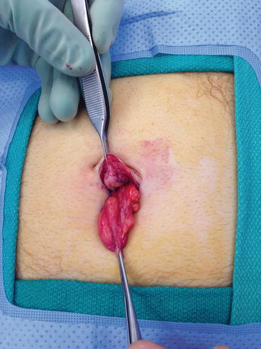
Figure 33.3
The hernia sac is dissected free of its attachments to the fascia and replaced within the abdomen
Primary Repair
For a primary repair, the fascial edges are dissected out and reapproximated with interrupted braided polyester sutures (Ethibond, Johnson & Johnson) (Fig. 33.4). This can be done with interrupted or figure of 8 type sutures or utilizing a vest over pants technique. The base of the umbilicus is resecured to the underlying fascia with a single simple absorbable suture, and the overlying skin is then closed. In order to prevent seroma formation we place a cotton ball in the umbilicus, cover it with an occlusive dressing, and aspirate the air with a needle to create a pressure dressing (Fig. 33.5).
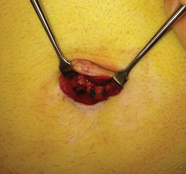
Figure 33.4


Simple interrupted sutures close the hernia defect in this primary repair
Related posts:


Stay updated, free articles. Join our Telegram channel
Full access? Get Clinical Tree
