Past Medical History:
Hypertension, COPD, obesity, smoker
Medications:
Lovastatin 40 mg oral daily
Amlodipine 10 mg daily
Valsartan 320 mg daily
Atenolol 25 mg daily
Allergies:
Iodinated contrast dye—anaphylaxis
Physical Exam:
VS:
BP 144/96 HR 77 RR 11 SaO2 98% (room air) Ht 70″ Wt 300 lb (136 kg)
General:
Obese, comfortable
Airway:
MP3, thick neck, large tongue, full neck extension and mouth opening, four finger thyromental distance
Chest:
Symmetric air entry bilaterally without crackles or wheezes
Data:
PFT: PreFEV1 47%, PreFVC 71%, DLCO 50%
Exercise treadmill myocardial perfusion imaging study:
Exercised for 7:00, MPHR 90%, RPP: 21,900, terminated due to dyspnea, 9 METS, non-ischemic, post stress LVEF 60%.
Chest CT:
5 × 4 cm mass obstructing the left lower lobe bronchus. Multiple 1 cm left hilar lymph nodes. Moderate to severe bilateral upper lobe predominant emphysema.
- 1.
What is the functional anatomy of the lungs?
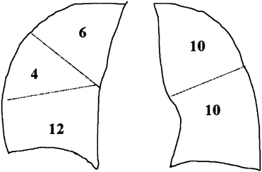
The trachea forms from the lower larynx at approximately C6, is approximately 15 cm (20–25 cm from teeth to carina) in an adult male, consists of 16 to 20 cartilaginous rings, lies directly anterior to the esophagus, and bifurcates at T4. The right mainstem bronchus is shorter compared with the left (i.e., 2.5 vs. 5 cm) and descends more vertically, thus the tendency for right mainstem intubations. The right lung consists of upper, middle, and lower lobes, with three, two, and five segments, respectively. The left lung consists of upper and lower lobe, each with five segments. Each pyramidal-shaped segment receives a single branch of the pulmonary artery, which follow along the bronchi and bronchioles to perfuse the alveoli. Smaller bronchial arteries supply the conducting airway, pleura, and nodes. For the purposes of predicting postoperative predicted FEV1 (ppoFEV1), a total of 42 lung subsegments are considered (RUL: 6, RML: 4, RLL: 12, LUL: 10, LLL: 10) (see Fig. 14.1).


Fig. 14.1
The number of subsegments of each lobe are used to calculate the predicted postoperative (ppo) pulmonary function. There are 6, 4, and 12 subsegments in the right upper, middle, and lower lobes. There are 10 subsegments in both the left upper and the lower lobes, for a total of 42 subsegments. Following removal of a functioning right lower lobe, a patient would be expected to lose 12/42 (29%) of their respiratory reserve. If the patient has a preoperative FEV1 (or DLCO) 70% of predicted, the patient would be expected to have a ppoFEV1 = 70% (1–29/100) = 50%. Reproduced from Slinger and Darling [2]
- 2.
What are the concerns in preoperative risk assessment for lung resection?
The “three legged stool” consists of (1) respiratory mechanics, (2) cardiopulmonary reserve, and (3) parenchymal function.
The main measure of respiratory mechanics is the FEV1. A ppoFEV1 can be estimated by multiplying the preoperative FEV1 (preFEV1) by the fraction of lung remaining at the conclusion of surgery. Respiratory complications are increased when ppoFEV1 < 40%, and <30% predicts high risk. In this patient, 10 of 42 subsegments will be resected, therefore


Cardiopulmonary reserve is formally described by maximum oxygen consumption and approximated by stair climbing (at patient’s own pace but without stopping) and 6 min walk. VO2max > 20 cc/kg/min predicts low risk and is approximated by ≥5 flights of stairs whereas the commonly used two flights of stairs correspond to a VO2max of 12 cc/kg/min. A ppoVO2max < 10 cc/kg/min predicts very high risk and is approximated by inability to climb 1 flight of stairs; this may be an absolute contradiction to lung resection. 1 MET corresponds to a VO2 of 3.5 cc/kg/min. In this patient:


Gas exchange is estimated by DLCO, PaO2, and PaCO2. ppoDLCO < 40% predicts increased respiratory and cardiac complications, and <20% is typically considered unacceptably high risk. PaO2 < 60 mmHg and PaCO2 > 45 mmHg are also indicators of increased risk. In this patient:


- 3.
What are the additional anesthetic considerations for lung cancer patients (the “four Ms”)?
Mass effects—is there compression of heart, airway, vein, artery, or nerve?
Metabolic—are there electrolyte abnormalities (hyponatremia, hypercalcemia), or paraneoplastic syndrome such as Lambert–Eaton?
Metastases—for example, are there brain or bone lesions that might change management?
Medications—have there been chemotherapy exposures, as to bleomycin or doxorubicin, which may change management or prompt further workup? [1]
- 4.
Will you advise this patient to quit smoking?
The answer should always be yes. The longer the cessation the greater the benefit, with perhaps 8 weeks being a worthwhile goal. However, even a 12 h cessation will decrease carboxyhemoglobin concentrations. And preoperative cessation predicts postoperative cessation, which has important implications for wound healing and infection.
- 5.
What are the indications for lung isolation? Does this case require lung isolation and OLV?
Indications for lung isolation may be absolute or relative. Absolute indications include for the prevention of spillage of pus, blood or large volume lavage fluid from the contralateral lung; bronchopleural fistula (in which the low resistance pathway can rapidly create pneumothorax during positive pressure ventilation); large unilateral bullae prone to rupture; and during video assisted thoracoscopic surgery (VATS) procedures. Relative indications include pneumonectomy, lobectomy (especially upper), thoracic aortic aneurysm repair, and esophageal surgery. While a relative indication, lung isolation during lobectomy will provide more ideal surgical conditions.
- 6.
How is lung isolation achieved? What are the advantages and disadvantages of the main methods of lung isolation?
Stay updated, free articles. Join our Telegram channel

Full access? Get Clinical Tree


