Fig. 14.1
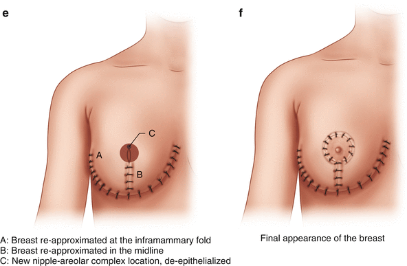
(a) Standard markings for oncoplastic surgery. (b) Standard wise pattern markings. (c) Isolating the inferior pedicle. (d) Creating the breast mound. (e) Relocation of the nipple-areolar complex. (f) Final appearance of the breast
(a)
Preoperative marking is imperative (Fig. 14.1a)
i.
Mark the sternal midline, the inframammary fold (IMF), the sternal notch to nipple distance, and the breast meridian in the sitting position. The breast meridian is the midpoint of the clavicle through the midline of the breast—this usually bisects the nipple-areolar complex. The new location of the nipple-areolar complex is based on the intersection of the breast meridian and the inframammary fold. Mark all distances.
(b)
With an inferior pedicle technique, the nipple is usually moved 8–10 cm cranially, and derives its blood supply from the breast tissue along the natural inframammary fold—the inferior pedicle. It is vital that this pedicle not be too narrow, or undercut in any way, as this could lead to nipple ischemia or loss. (Fig. 14.1b)
(c)
Inscribe the nipple-areolar complex with a round metal “cookie cutter,” aiming for a 4 cm diameter in most reductions, at the level previously marked and measured in pre-op (Fig. 14.1b).
(d)
Incise the pedicle along the markings, and de-epithelialize from the nipple to the inframammary fold, taking care to preserve the subdermal plexus. (Fig. 14.1c)
(e)
Flaps of at least 1 cm in thickness are developed up to the clavicle with electrocautery. Several large vessels are usually encountered and addressed with targeted electrocautery.
(f)
The medial and lateral margins of the inferior pedicle are defined with electrocautery, and then sharply divided with a Watson blade (taking care not to bevel into the pedicle) down to the chest wall. This enables all tissue medial and lateral to the pedicle (below the inframammary fold) to be resected. With an inferior pedicle, the tissue resected is usually superior and lateral. Take care not to apply tension or torque to the pedicle. Preserve pectoral attachments within the pedicle. (Fig. 14.1c)
(g)
Place a 0-silk suture through the nipple-areolar complex (in situ on the inferior pedicle).
(h)
Breast parenchyma is re-approximated, starting with a buried 3-0 absorbable suture placed to define the new midpoint of the inframammary fold—the “triangle corner stitch”—to incorporate the medial and lateral flaps with the center of the inframammary fold. (Fig. 14.1d)
(i)
Close the remaining dermal layers with interrupted buried 3-0 absorbable suture.
(k)
Remove any dermal sutures needed to allow for use of the previously placed 0-silk suture to deliver the nipple into the new location (previously delineated with the “cookie cutter”). (Fig. 14.1e)
(l)
Close the entire incision with a running 4-0 monofilament suture.
(m)
Tack the areola in place using buried interrupted 3-0 absorbable suture, placed first at the cardinal points, and then with sutures in between, until the areola is secure.
(o)
All tissue from any breast reduction should be sent for pathologic evaluation. Drains may be left at the discretion of the surgeon.
2.
Get Clinical Tree app for offline access

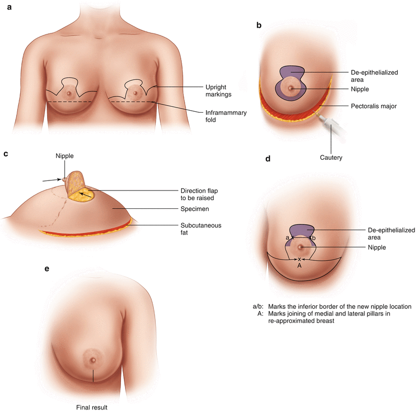
Retro-areolar or lower outer/lower inner quadrant tumors (Fig. 14.2):


Fig. 14.2


(a) Standard markings for oncoplastic approach to lower quadrant tumors. (b) Mobilization of the specimen. (c) Creation of the nipple flap. (d) Re-approximation of the breast. (e) Final appearance of the breast
Stay updated, free articles. Join our Telegram channel

Full access? Get Clinical Tree


