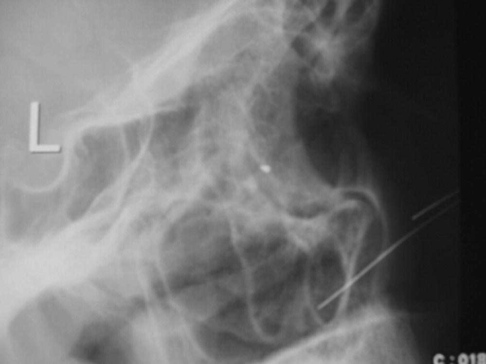
Chapter 37 Eric Hawkins and Michael D. Mills Traumatic eye injuries are extremely common in the prehospital setting, and may occur as isolated injuries or as part of more extensive maxillofacial or multisystem trauma. These injuries may range from the minor to the sight threatening, and EMS physicians must be prepared to rapidly identify serious problems that could result in permanent blindness or further complications. Once significant eye injuries are recognized, it is important that the patient is stabilized, appropriately treated, and evaluated by a hospital or physician with adequate access to full ophthalmological services to provide definitive care. Ocular trauma is common; in the United States an estimated 2–3 million people seek medical attention for eye injuries each year [1,2]. Among many risk factors, the most significant seem to be male sex and age under 30 [3]. Most injuries are not significant, and many never need treatment for minor eye problems [2]. Of those with more serious injuries, 16% have ocular or orbital damage and over 50% of patients with significant facial trauma have associated sight-threatening eye injuries [4]. Trauma is the second most common cause of monocular blindness, trailing only cataracts [5]. Each year, eye injuries are the number 1 cause of ophthalmological hospital admissions in the United States [5]. Initial assessment and treatment should focus on the ABCs of trauma resuscitation, and any life-threatening injuries should be addressed first, as with any trauma patient [6,7]. Eye injuries can be distracting, and it is important not to divert attention from other sources of serious injury early in the trauma survey process. It is also critical to recognize that associated facial trauma and swelling may affect airway patency, and this should be secured before further examination of the orbit if needed. After initial stabilization and primary survey, attention can be focused on the ocular injury and a thorough evaluation should be performed. In the case of known or suspected chemical contact to the face and eye, immediate irrigation with normal saline or clean water should be performed during this evaluation process The key components in the evaluation of traumatic eye injuries are a thorough history and careful eye examination. The history focuses on key points surrounding the event and should note the type of injury, the time of onset, and any specific symptoms reported by the patient [6]. Mechanism of injury is also recorded and may include blunt or penetrating trauma and thermal or chemical burns to the eye or periorbital areas of the face. Other important points include the patient’s visual acuity before the injury, if known, the presence or absence of contact lenses, any past medical history of eye disorders, and any history of ophthalmological surgical or medical treatment [7]. The physical examination of the eye begins with evaluation of visual acuity, establishing a baseline level of function and providing functional assessment of possible damage to the eye [7,8]. In the field, this can be performed using a hand-held Snellen chart to document the smallest objects or letters identifiable at a specific distance from the eye. Visual acuity is recorded for each eye individually and then using both eyes simultaneously [7]. If no chart is available, a newspaper or other source of small print is useful to estimate visual acuity. Patients who normally wear prescription glasses for reading should perform this with those same glasses if available, but those who use contact lenses should not have those replaced for this examination. If the patient’s glasses are unavailable, it is possible to use a piece of paper with a small pin-sized hole through which the patient can view the chart and complete the examination [6,7]. This “pin-hole test” corrects for the refractive error of the patient’s eyes and should allow completion of the examination. For those who cannot read the Snellen chart due to injury or underlying ocular disease, other options include assessing the patient’s ability to count fingers, detect hand motions, or perceive the presence or movement of light [6]. The method of testing and patient performance should be documented for each eye. After rapid evaluation of visual acuity, attention shifts to the external assessment of the eye and surrounding structures. Each globe is examined for protrusion or proptosis and for external signs of penetration or damage from a foreign body (Figures 37.1–37.3). Ocular movement in the cardinal directions of gaze (vertical up-down, horizontal right-to-left, and diagonal left-to-right and right-to-left) is also tested and any deficit or entrapment recorded. The pupil and iris are then inspected for size, shape, and reaction to light and results compared between eyes. The presence or absence of a hyphema (blood in the anterior chamber that may obscure the iris or pupil) is especially important to assess (Figure 37.4). Finally, the conjunctivae are inspected for erythema, subconjunctival hemorrhage, chemosis, conjunctival swelling, or subconjunctival emphysema. If the patient reports a foreign body sensation in the eyelid (Figure 37.5) or if there is any concern for an intraocular foreign body or a punctured globe (Figure 37.6), it is best to end the examination at this point. The affected eye should then be covered with an eye shield or improvised protective device to protect the globe from external pressure before transport for more definitive evaluation and care [7]. The EMS physician should not remove a protruding foreign body (such as a nail) lodged in the globe. A cup or shield may be used to cover the eye with the foreign body in place.
Ocular trauma
Introduction
Epidemiology
Evaluation: history and physical exam


Stay updated, free articles. Join our Telegram channel

Full access? Get Clinical Tree


