(1)
Critical Care Medicine and Pain Medicine, Boston Children’s Hospital, Boston, MA, USA
(2)
Harvard Medical School, Boston, MA, USA
Keywords
Pyloric stenosisEsophageal atresiaGastroschisisOmphaloceleCongenital diaphragmatic herniaSacrococcygeal teratomaCongenital lobar emphysemaVATER associationA 14-hr-old male, 2,400 g, born at 37 weeks’ gestational age, is scheduled emergently for repair of an esophageal atresia with tracheoesophageal fistula. The newborn choked and gagged on the first glucose water feed. A contrast study confirmed the diagnosis. An NG tube is in place. The infant is receiving nasal cannula oxygen at 300 mL/min.
VS: HR = 158/min; BP = 88/52 mmHg; RR = 44/min; T = 37.2 °C. SpO = 95 %; Hgb = 13.0 g/dl.
Preoperative Evaluation
Questions
- 1.
Is this an emergency? What historical information might have helped anticipate the diagnosis? Are there other, safer, means to make the diagnosis than a contrast study?
- 2.
What other evaluations of the newborn are needed prior to undertaking the induction of anesthesia? What conditions are likely or associated with EA/TEF?
- 3.
Is a preoperative gastrostomy with local anesthesia indicated? Would the situation be different if the patient were preterm with respiratory distress syndrome (RDS)?
Preoperative Evaluation
Answers
- 1.
While repair of the esophageal atresia (EA) with tracheoesophageal fistula (TEF) may not be a true emergency, it is, at the least, very urgent. The longer the newborn is unrepaired, the greater the risk for aspiration. Surgical correction should proceed very quickly but proper preparation can be accomplished in short order. The diagnosis can be suspected in cases of maternal polyhydramnios. In the delivery room, inability to pass a suction catheter into the stomach should raise the suspicion of EA. A contrast study is not needed to make the diagnosis. Aspiration of oral contrast is a significant risk. Plain X-rays may show the dilated, air-filled esophageal pouch. A film with a radiopaque catheter coiled in that pouch will confirm the diagnosis. If there is no gas in the abdomen, it is possible that the child has EA without TEF.
- 2.
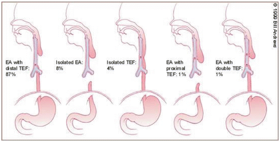
It is important to ascertain which type of TEF is present. In cases of esophageal atresia, >90 % have an associated tracheoesophageal fistula. The most common variant of a TEF, by far (90 %), is esophageal atresia with a distal fistula between the posterior trachea near the carina and the stomach. The next most common, approximately 7–8 %, is EA without TEF. Many other types and subtypes have been described. Up to 50 % of patients with EA/TEF have other congenital anomalies. Cardiovascular anomalies make up one-third of the anomalies seen in these patients. The cardiac anomalies seen are, in order of occurrence, VSD, ASD, tetralogy of Fallot, and coarctation of the aorta. Other organ systems involved in these patients are musculoskeletal (30 %), gastrointestinal (20 %), and GU (10 %). Patients with EA/TEF may have the VATER syndrome that consists of vertebral defects or VSD, anal/arterial defects, TEF/EA, and radial or renal anomalies [2, 3].

- 3.
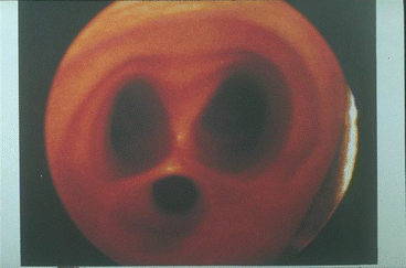
There are three methods used to decrease or eliminate insufflation of the stomach with the inspired gas from the endotracheal tube. The endotracheal tube tip can be placed beyond the fistula, just above the carina, but in some cases the fistula is actually at the carina making this procedure impossible. In cases where the newborn is having severe respiratory compromise and positive pressure ventilation has been instituted, a ventilator breath may follow a path from the trachea through the fistula and distend the stomach. The abdomen can become very distended, further compromising ventilation. In these dire situations, an emergent gastrostomy may allow the abdominal pressure to be relieved enough for ventilation to continue [4]. Approximately 25 % of newborns with EA/TEF are born preterm, and in cases with respiratory distress, the situation is even more difficult since institution of positive pressure ventilation will require higher pressures. This will invariably also put gas into the stomach through the fistula. In cases when ventilation of the lungs is ineffective or incomplete, another option in addition to an emergency gastrostomy is placement of a balloon-tipped catheter through the fistula into the stomach, inflating the balloon and occluding the fistula. This can be accomplished by placing the balloon-tipped catheter through the fistula from the trachea with a rigid bronchoscope. Photo below shows the relative sizes of the right and left bronchi and the fistula with a positive pressure breath. Spontaneous breathing may not illustrate this as well, under direct vision.


Intraoperative Course
Questions
- 1.
What monitors would you require for this case? Arterial line? Where? How will you assess intravascular volume? CVP catheter? How much information about preload would a Foley catheter give you for this case?
- 2.
What might be done to minimize the effect of the fistula at induction? Is IV or inhalation induction preferable? How should the airway be secured? Does the presence of the fistula alter the techniques for intubation? What should be done with the NG tube once intubation is accomplished?
- 3.
Is controlled or spontaneous ventilation preferable for these cases? How would you determine whether or not a percutaneous gastrostomy is indicated prior to the definitive repair? Is a precordial stethoscope of particular importance for these cases?
- 4.
After positioning and the start of the thoracotomy, breath sounds from the left axillary stethoscope markedly diminish and the SpO2 decreases. What might be the cause? What would you do? Could the endotracheal tube have accidentally entered the fistula?
Intraoperative Course
Answers
- 1.
For otherwise well term newborns with EA/TEF, standard monitors, with the addition of “pre-” (right hand) and “post-” (left hand or either foot) ductal pulse oximeters and a Foley catheter, will often be sufficient. If there is pulmonary compromise, either from aspiration or because of prematurity, an arterial line is useful for frequent ABG determinations. A CVP catheter would not only give some information about intravascular volume but also be an excellent route for administration of resuscitation medications, should that be needed. If peripheral IV access is good in an otherwise well newborn with EA/TEF, the risk of placing a CVP line may not be justified. Urine output should mirror renal blood flow (GFR) but it is a secondary measure. In addition, the small volume produced may be difficult to accurately collect and measure. Nevertheless, this monitor can provide useful information for these cases.
- 2.
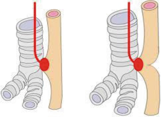
In cases where the connection between the trachea and esophageal point if entry of the fistula is open, avoidance of positive pressure ventilation is important. Positive pressure ventilation will force gas through the fistula into the stomach. IV access should be secured prior to any attempts at induction of anesthesia or intubation. Awake intubation is often done, followed by spontaneous ventilation with the infant breathing oxygen plus incremental doses of a volatile anesthetic. Alternatively, an inhalation induction can be done, and when an adequate depth of anesthesia has been achieved and the airway anesthetized with the appropriate dose of topical anesthetic, laryngoscopy and intubation can be done. It has been commented that turning the bevel of the endotracheal tube anteriorly will decrease the chance of intubating the fistula but this is unproven. It also has been suggested that since the fistula is often relatively low in the trachea, a deliberate right main stem intubation should be done and the endotracheal tube then withdrawn to a position just above the carina, hopefully distal to the fistula. Great care is required while advancing the endotracheal tube in the trachea, however. The fistula may be quite large and the endotracheal tube may easily be placed into the fistula if it is advanced too far into the trachea [5]. As mentioned above, an alternative that will allow positive pressure ventilation is performance of a rigid bronchoscopy following induction of anesthesia and placement of an occluding balloon-tipped catheter through the fistula. The balloon is then inflated and the catheter pulled taut, thus closing the fistula [6]. This technique allows positive pressure ventilation to proceed without distending the stomach. The surgeon will likely ask that the NG tube be advanced during the procedure to facilitate identification of the esophageal pouch.
- 3.
If the stomach distends after intubation, even with gentle assistance of respiratory efforts, and this distention is interfering with ventilation (leading to the use of higher ventilation pressures), percutaneous gastrostomy will allow some control of the situation. The usual position for surgery is left side down for a right thoracotomy. The surgeon retracts the right lung, leaving only the left lung for gas exchange. In this situation, a left axillary stethoscope will give the anesthesiologist immediate information about the adequacy of ventilation. The left bronchus is easily occluded by blood or secretions and may be kinked by the surgeon during the procedure; the anesthesiologist must be aware of these events as soon as they occur [7].
- 4.
Secretions and/or blood may easily occlude the lumen of the trachea or right main bronchus. Additionally, the bronchus is often kinked by surgical retraction during the procedure. Even with occlusion of the fistula by a balloon-tipped catheter, given the relatively large size of some fistulae as seen in the photo above, there still may be room for the tip of the endotracheal tube to completely or partially enter the fistula, greatly decreasing or eliminating ventilation of the lungs [7].
Stay updated, free articles. Join our Telegram channel

Full access? Get Clinical Tree


